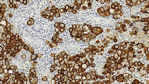
Human skin, melanoma: immunohistochemical staining for HMB45. Note the melanoma showing strong cytoplasmic staining. Melanoma Marker (HMB45): clone HMB45
Melanoma Marker (HMB45)
Antigen Background
The HMB45 antigen has also been identified in retinal pigment epithelium (RPE) but is reported to be reactive only with the transient prenatal and infantile RPE. No reaction is reported to be observed with intradermal nevi and normal adult melanocytes and non-melanocytic cells.
Tumor cells of epithelial, lymphoid, glial and mesenchymal origin are reported to be negative. This clone is well described in the literature. It is indicated to label an intracytoplasmic antigen in the majority of melanomas and other tumors demonstrating melanoma/melanocytic differentiation.
Product Specific Information
The clone is also reported to react with junctional and blue nevus cells. (Bacchi CE et al., A Review. Applied Immunohistochemistry. 4:73-85 (1996)).
Disclaimer
Melanoma Marker (HMB45) is recommended for the detection of specific antigens of interest in normal and neoplastic tissues, as an adjunct to conventional histopathology using non-immunologic histochemical stains