-
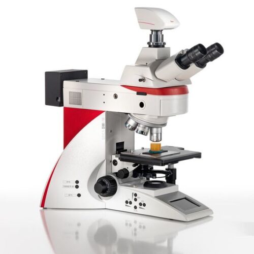
 The Leica DM2700 M flexible upright microscope system uses LED illumination for all contrast methods: brightfield (BF), darkfield (DF), differential interference contrast (DIC), qualitative polarization (POL), and fluorescence (FLUO) applications. It also offers built-in oblique illumination, which improves the visualization of surface topography and defects. Optionally, the Leica DM2700 M can also be equipped with a transmitted light axis. The Leica DM2700 M is equipped, e.g. with an N PLAN achromatic objective series with magnifications from 5x to 100x, a field of view of 22 mm, a flattened image field, and large working distances.
 -
The Leica DM2700 M flexible upright microscope system uses LED illumination for all contrast methods: brightfield (BF), darkfield (DF), differential interference contrast (DIC), qualitative polarization (POL), and fluorescence (FLUO) applications. It also offers built-in oblique illumination, which improves the visualization of surface topography and defects. Optionally, the Leica DM2700 M can also be equipped with a transmitted light axis. The Leica DM2700 M is equipped, e.g. with an N PLAN achromatic objective series with magnifications from 5x to 100x, a field of view of 22 mm, a flattened image field, and large working distances.
-
 The Leica DM2700 M flexible upright microscope system uses LED illumination for all contrast methods: brightfield (BF), darkfield (DF), differential interference contrast (DIC), qualitative polarization (POL), and fluorescence (FLUO) applications. It also offers built-in oblique illumination, which improves the visualization of surface topography and defects. Optionally, the Leica DM2700 M can also be equipped with a transmitted light axis. The Leica DM2700 M is equipped, e.g. with an N PLAN achromatic objective series with magnifications from 5x to 100x, a field of view of 22 mm, a flattened image field, and large working distances.
 -
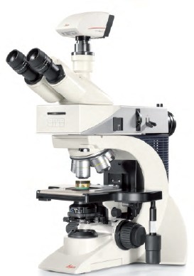
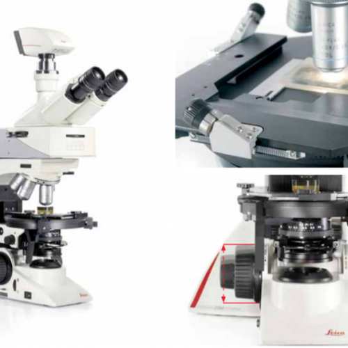
 Depending on your application, you can choose from three different systems for polarization microscope. The Leica polarization microscope series is designed for all polarizing examinations: petrography, mineralogy, structure characterization, asbestos analysis, coal analysis (vitrinite reflection), and examination of liquid crystals.
 -
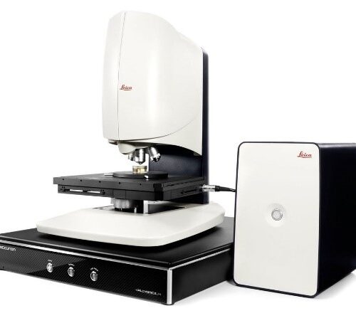 Designed to help you maximize efficiency, Leica DCM8 unites the advantages of High Definition confocal microscopy with interferometry into one versatile, dual-core system. Ultra-fast analysis is ensured thanks to one-click mode selection, sophisticated software and HD confocal scanning without moving parts.
 -
 System for Microelectronics and Semiconductor. With a large field of view, the DM3 XL inspection system allows your team to identify defects faster and increase your yield rate. Make use of the 30% increased field of view of the unique macro objective. The DM3 XL uses LED illumination for all contrast methods. LED illumination provides a constant color temperature and offers real color imaging at all intensity levels.
 -
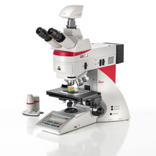
 The integrated laser spectroscopy function of the DM6 M LIBS delivers the chemical composition of the microstructure that you see in the microscope image - within a second. Identify the microstructure composition of interest, then trigger the LIBS analysis with a single click. The DM6 M LIBS 2-in-1 solution allows you to perform both structural and elemental/chemical analysis of material phases, e.g., minerals, alloys, ceramics, etc. There is no need for sample preparation nor transfer between 2 or more devices. The entire analysis workflow occurs with a single instrument.
 -
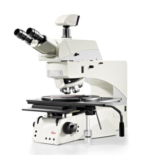 The Leica DM8000 M and Leica DM12000 M optical inspection systems provide an innovative yet cost-effective system solution for mastering present and future inspection challenges with confidence. Inspection, process control and defect analysis of wafers or LCDs and TFTs has to be fast, To detect macro defects, the Leica DM8000 M and DM12000 M have a micro/macro mode for rapid scanning of large components. The macro magnification captures an object field of approximately 40 mm.
 -
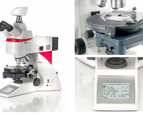
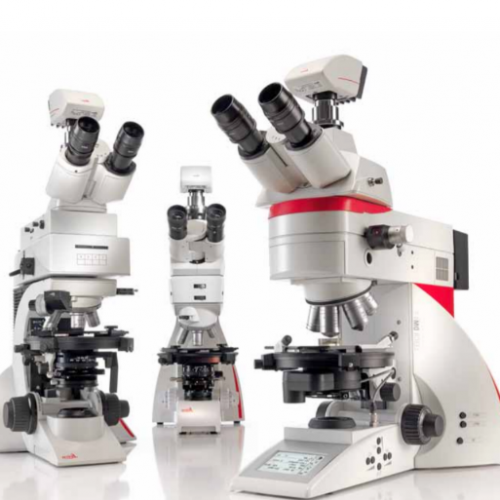
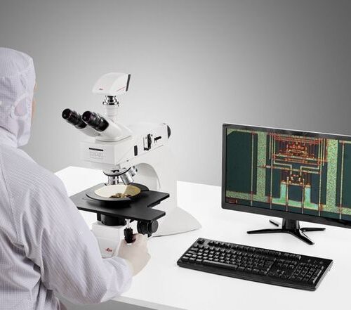
 Leica DM4 M and DM6 M digital microscopes for materials science and quality control offer truly reproducible microscopy, incredible optics and high-quality images. Store and recall your imaging conditions with a touch of a button. High quality microscope images make challenging inspection, measurement, and analysis tasks easy. Use the Leica DM4 M for manual routine inspection or the Leica DM6 M for fully automated materials analysis.
 -
 The Leica DM2700 M flexible upright microscope system uses LED illumination for all contrast methods: brightfield (BF), darkfield (DF), differential interference contrast (DIC), qualitative polarization (POL), and fluorescence (FLUO) applications. It also offers built-in oblique illumination, which improves the visualization of surface topography and defects. Optionally, the Leica DM2700 M can also be equipped with a transmitted light axis. The Leica DM2700 M is equipped, e.g. with an N PLAN achromatic objective series with magnifications from 5x to 100x, a field of view of 22 mm, a flattened image field, and large working distances.
 -
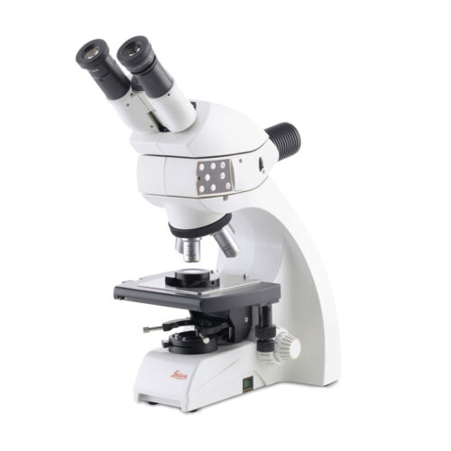 The Leica DM750 M is the ideal microscope for basic materials applications in an Industrial Lab or Material Science course. It can be equipped with various specimen holders to accommodate mounted specimens of different diameters. The unique Reflected light LED illuminator provides brightfield, polarized light, and oblique illumination. This allows you to work with many different specimens with the same microscope configuration.