Antigen Background
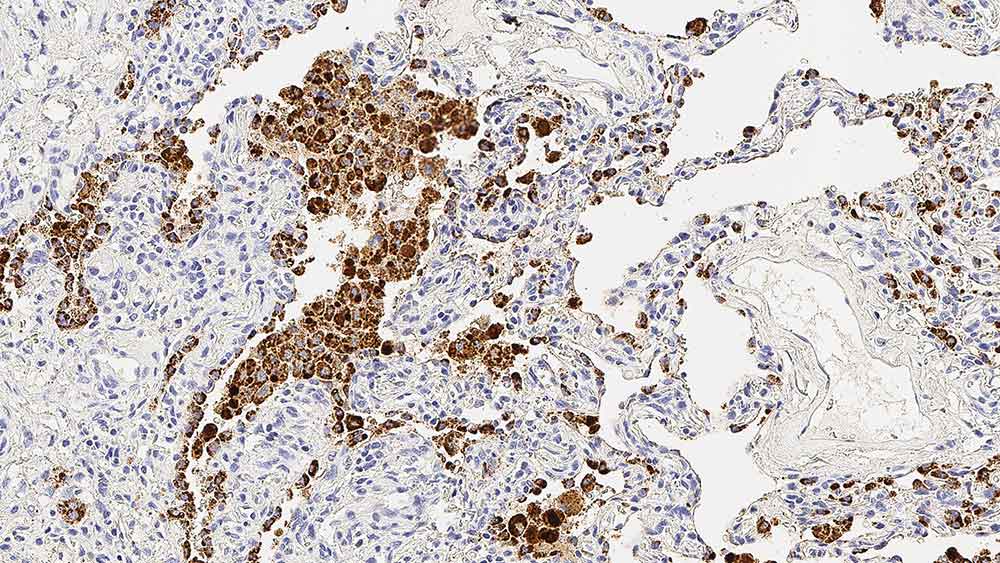
Napsin A has a specific function in normal alveolar epithelium and is proposed to play a role in the proteolytic processing of surfactant precursors.
Napsin A is reported to be predominantly expressed in lamellar bodies of type II pneumocytes, secondary lysosomes of alveolar macrophages, respiratory epithelium of terminal and respiratory bronchioles, plasma cells, within a subset of lymphocytes in normal lung, as well as in epithelial cells of renal tubules in normal kidney and is weakly expressed in normal spleen.
Studies have reported that Napsin A is expressed in 90% of primary lung adenocarcinomas.
Disclaimer
Napsin A is recommended for the detection of specific antigens of interest in normal and neoplastic tissues, as an adjunct to conventional histopathology using non-immunologic histochemical stains.