See more of the surgical site in one view so you can progress seamlessly through your next procedure with the PROvido floor stand surgical microscope.
PROvido provides you with a bright, fully-focused view deeper into narrow cavities. A challenge that occurs daily in otolaryngology and spine procedures.
PROvido
To view pricing, add to cart and log in.
Contact Us for a quote
Contact Us for a quote
Description
See more, simply.
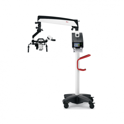
Multidisciplinary Surgical Microscope PROvido
See more of the surgical site in one view so you can progress seamlessly through your next procedure with the PROvido floor stand surgical microscope.
PROvido provides you with a bright, fully-focused view deeper into narrow cavities. A challenge that occurs daily in otolaryngology and spine procedures. With exclusive Fusion Optics technology and concentrated xenon light PROvido unites advantages of premium surgical optics for the first time with a responsive, stable floor stand.
No need to search for vital details or constantly refocus. Work smoothly and confidently throughout your procedure with the PROvido multidisciplinary microscope that allows you to see more, simply.
Always focused on your patient Avoid constantly interrupting surgery to refocus thanks to FusionOptics which provides a high resolution view to the bottom of deep cavities. Get into the position and get going Set up quickly and effortlessly, and be sure the microscope remains exactly where you need it due to the lightweight, robust design. Proceed without limitation Enjoy the comfort and workflow benefits of adaptable optics and more working space, even when working with long instruments.
The Technology of FusionOptics
- Two separate beam paths
- One beam path provides high resolution.
- The other beam path provides a depth of field.
- The brain merges the two images to a single optimal spatial image.
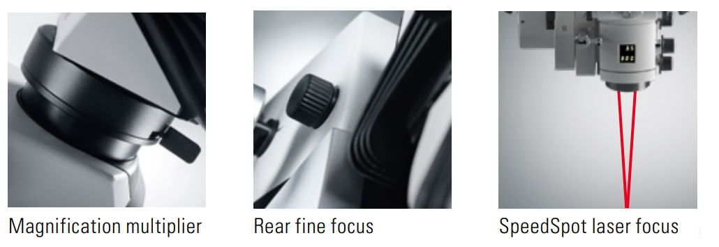
The view you need, fast. Meet the visualization needs of your procedure and your team at every moment with optics that are fast to set up and adjust. > Start fast with the SpeedSpot system that uses two laser beams as a focusing reference to quickly provide a defined focus point for all three viewing positions (surgeon, assistant, camera) > Quickly achieve maximum magnfication with the optional Magnification Multiplier that boosts magnification by 40% in an instant > Give your opposite assistant viewing flexibility with an independent fine focus.
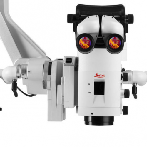
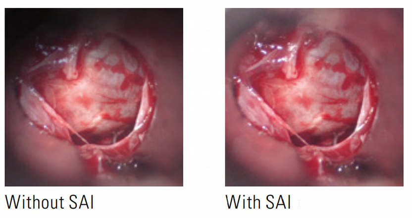
Deeper insights Small Angle Illumination (SAI) technology and bright 300-Watt xenon light provides a concentrated light beam. You benefit from a bright view with less shadow right down to the bottom of deep, narrow cavities.
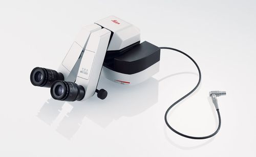
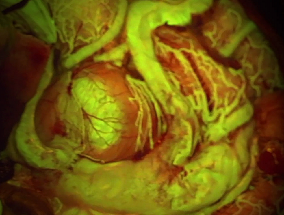
FL580 Fluo Module, Simplify your workflow
Simultaneous anatomical and real-time fluorescence visualization means no need to interrupt workflow to switch back and forth between views.
Thanks to full microscope integration, activating FL560 mode requires just one click of the handgrip or footswitch. If you choose to integrate other fluorescence modes, you can also switch between them with a single click.
Simply activate FL560 mode and keep working!
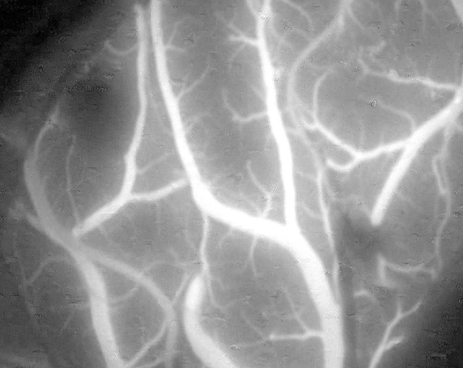
Intra-Operative Fluorescence-Guided Blood Flow View 820 nm/NIR Leica FL800
A ruptured cerebral aneurysm is the third most common cause of death in industrial countries.
With the Leica FL800, for intra-operative fluorescence-aided video angiography, surgeons can now determine the patency of vessels.
During surgery, the patient is injected intravenously with ICG (indocyanine green) dye, which is well tolerated and spreads quickly. A near-infrared camera then shows the bloodstream in black-and-white directly through the surgical microscope eyepieces and/or on a video monitor.
OPTICS AND ILLUMINATION
FusionOptics For increased depth of field and high
resolution for the main surgeon
Fully apochromatic
optics
For high contrast and natural colours
without chromatic aberrations
Magnification 6:1 zoom, motorized
Optional 1.4× magnification multiplier
Focus Motorized via a multifocal lens, with
manual adjustment
Objective /
working distance
225–600 mm, motorized multifocal lens,
continuously adjustable and manual
adjustment option
Eyepieces Wide-field eyepieces for persons wearing
glasses 8.3×, 10× dioptric adjustment, ±5
diopter settings and adjustable eyecup
Integrated 360°
rotatable adapter
For main surgeon binocular (IVA, ULT) and
opposite assistant (ULT)
Illumination – Single Xenon 300-Watt arc-lamp as
main illumination with LED lamp as
back up or optional dual Xenon arclamp
illumination system
– Continuously variable illumination field
diameter
– Continuously adjustable brightness at
constant colour temperature
SpeedSpot Laser focusing aid for fast and exact
the positioning of the microscope
AutoIris Built-in automatic zoom-synchronized
illumination field diameter, with manual
override and reset feature
BrightCare Plus Safety function through working distance dependent
limitation of the brightness,
controlled by a built-in lux meter
CONTROL
Control unit – Programmable touch-screen with user friendly
Graphical User Interface for
control of the microscope and stand
– Built-in electronic auto-diagnosis and
user support
– Software independent hard keys for
illumination
– Indicator for main/backup illumination
and fluorescence modes
Control elements – Pistol handle with 10 programmable
functions
– Optional 12-function wireless
footswitch
IR sensor For remote control of the external Leica
HD C100 camera
MANEUVERABILITY
XY speed Zoom linked XY speed
XY range 62 x 62 mm
Balancing Manual balancing at microscope carrier
and at swing arm
Brakes Floor stand with 6 electromagnetic
brakes
Carrier for monitor 610 mm long and flexible arm with 180°
rotation and inclination to carry optional
video monitor
OPTIONS
ULT530 – Full stereo view for the main surgeon and
opposite assistant, semi stereoview
for 2 side assistants
– Optional integrated HD Camera
(HD C100)
FL800 ULT ULT with the Leica FL800 vascular
fluorescence observation filter module
FL560 Leica FL560 fluorescence observation
filter module
IVA530 – Full stereo view for the main surgeon,
semi stereo view for 2 side assistants
and C-mount interface for camera
(HD or SD)
Integrated documentation
Prepared for the integration of video camera
system and digital recording. Open
architecture
Digital Recording Record still pictures and videos in
standard-definition (SD) and high
definition (HD) using a digital recording
device (documentation system)
Laser compatibility
compatible with Lumenis AcuSpot 712L
Laser Manipulator and Digital AcuBlade
CONSTRUCTION
Base 700 × 700 mm with four 360° rotating
castors with a diameter of 126 mm each
and integrated brakes
Materials All solid metal construction
Load Max. 8.5 kg from microscope dovetail ring
Interface
Weight Approx. 350 kg without load