-
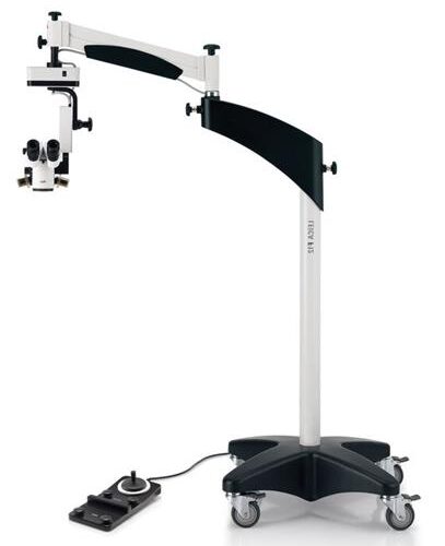
 The Leica M620 F20 – an ophthalmic surgical microscope with its crisp, sharp OptiChrome™ optics gives the surgeon natural color, outstanding depth-of-focus and higher contrast for maximum detail recognition.
 -
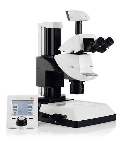
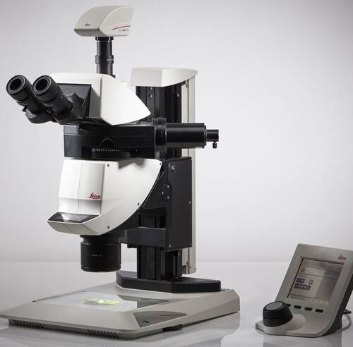
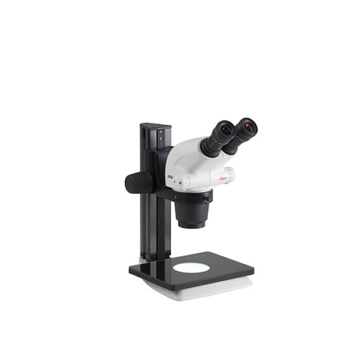
 M60 with 6:1-zoom range, with a continuous zoom from 6.3x – 40x including engageable click stops - making repetitive inspection and measurement at a specific zoom position reliable. This system is optimized for a larger field of view to see more at a glance.
 -
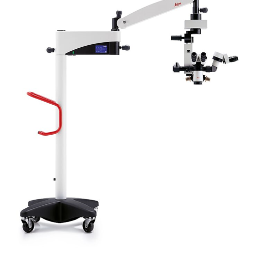
Precision Surgical Microscope For Neurosurgery, Spine & Plastic Reconstructive Surgery
Precision comes standard in our M530 OHX, helping you achieve the highest standards of precision in your surgery. -
You are always striving to reach the best possible patient outcome. To achieve this you need to see every detail even in narrow channels and to free maneuver yourself and your instruments.
-
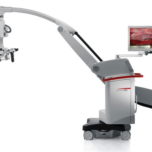
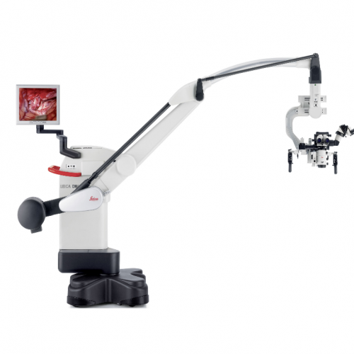 The Leica OH4 surgical microscope provides superior reach, height, and clearance. The Leica OH4 floor stands that moves easily and when the brakes are released, the microscope stabilizes in only 1.5 seconds and remains vibration-free. By integrating new glass, glass coatings, and design parameters, high-resolution APO OptiChrome technology delivers an expanded working distance and greater depth of focus. In combination with the innovative light management systems BrightCare and AutoIris, you can operate and safe light levels and still see more than ever before. The M525 OH4 also offers FL800 and FL400 fluorescence for intraoperative visualization of blood flow and tumour tissue.
 -
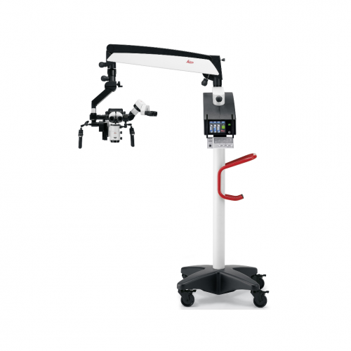
Compact surgical microscope Smooth, fine movements, Xenon illumination systems, Motorized MultiFoc lens
The Leica M525 F20 surgical microscope combines brilliant optical quality with superior maneuverability. The crisp, sharp images and large depth of field allow the surgeon to see precise details. The working-distance-based illumination provides enough light for deep operative sites while supporting patient safety. Designed as an otolaryngology microscope, the Leica M525 F20 is also ideal for spine, dental, hand, and plastic/reconstructive procedures. Ease of movement -
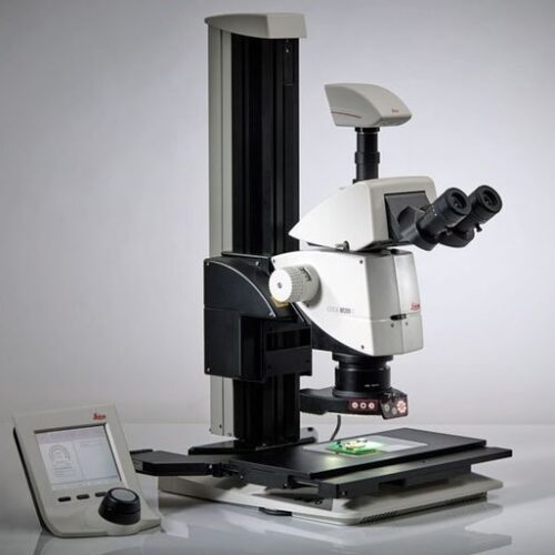
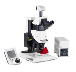
 with 5:1-zoom, featuring a step-zoom with five fixed zoom steps, at 6.3x, 10x, 16x, 25x, and 40x– allowing 100% reproducible results, this ensures that the results remain comparable at all times without great effort. The short optics carrier enables high light throughput and provides a large working distance, ideal in animal surgery and similar applications.
 -
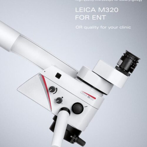
The Leica M320 F12 bundles apochromatic optical quality with cutting-edge LED illumination and high-definition imaging technology. Ease of use and flexible, smooth maneuverability are additional characteristics of this microscope.
-
 The Leica M220 F12 - an ophthalmic microscope is fully dedicated to the needs of ophthalmic surgery. Prestigious Leica optics motorized 5-step APO-chromatic magnification changer and focus, LED-illumination without fiber optics cables for direct and instant Red Reflex, and upgradeable XY-unit are standard features
 -
 Imagine not having to choose between high resolution or better depth of field, but to have both! The revolutionary FusionOptics™ technology makes it possible and provides you an ideal 3D image to see even the smallest details.
 -
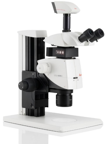
 The Leica M205 C, the world’s first stereomicroscope to offer 20.5:1 zoom, uses unique FusionOptics™ technology to provide exceptional image detail and impressive 3D depth perception. Its rigid, sturdy mechanical structure, revolutionary FusionOptics™, and completely integrated multi-contrast illumination, make the M205 C an ideal instrument for questioned document examiners seeking a high-performance microscope capable of revealing fine details.
 -
 Until now, you probably had to switch between two different systems: one for fast screening with a manual zoom that is intuitive to maneuver and a high-end solution to see and capture the faintest signals in an image.
 -
 Do you sometimes feel like you are fishing in the dark because you are looking for fluorescence signals that are increasingly faint and weak? Brightness and high resolution images are a must in modern developmental biology and research.
 -
 The Leica M205 FA opens a new world of research in fluorescence microscopy, for instance when working in a sterile cabinet.
 -
 The Leica M165 FC fully apochromatic corrected stereo microscope with 16.5:1 zoom optics resolves structures down to 1.1 micron for detailed fluorescent imaging.
 -
 The Leica M165 FC fully apochromatic corrected stereo microscope with 16.5:1 zoom optics resolves structures down to 1.1 micron for detailed fluorescent imaging.
 -
 Do you have to perform a wide variety of tasks but don’t want to constantly change the objective or even switch the microscope? Offering a large zoom and magnification range the Leica M165 C allows you manage all different kind of samples.
 -
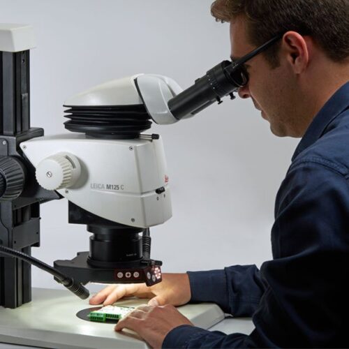
 With a coded zoom and apochromatic optics, the Leica M125 C offers high-end quality for mid-range budgets. Combining optimal resolution and depth of field with the integrated double iris diaphragm.
 -
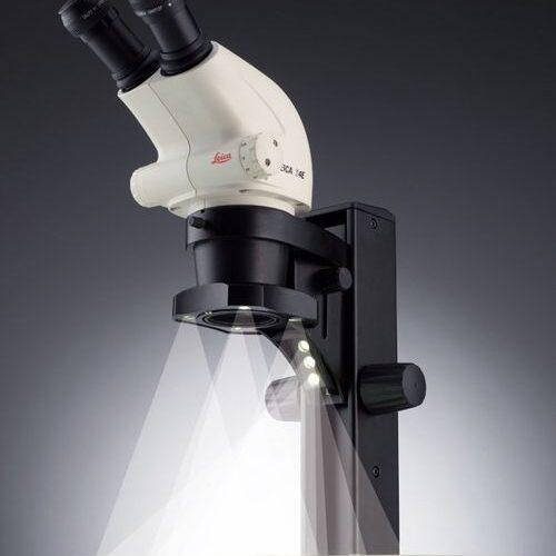
 With a Leica M-series stereo microscope you can choose from a comprehensive range of accessories to adapt your microscope to your application, whether it is in materials testing, life science research or forensic application.
 -
 With a Leica M-series stereo microscope you can choose from a comprehensive range of accessories to adapt your microscope to your application, whether it is in materials testing, life science research or forensic application
 -
 Unit Size 50 Each
 -
Unit Size 500 Each
-
 Unit Size: Box with 50 Each
 -
Unit Size: Case with 500 Each
-
 The Leica LED5000 CXI coaxial illuminator is used for quality control of flat, polished, or reflective samples. Scratches, cracks, impurities, and pores are made exceptionally visible. With coaxial illumination, light is coupled directly into a beam path and reflected into another beam path by the flat sample.
 -
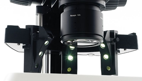
 The Leica LED3000 NVI is ideal for viewing recesses and holes, since the light falls almost vertically on the sample. In contrast to coaxial illumination it is also ideally suited for non-reflective and uneven sample Surface. NVI illumination shines almost straight down onto the sample plane. This allows cavities and recesses to be very well illuminated. The Leica NVI is installed between the microscope carrier and the objectives
 -
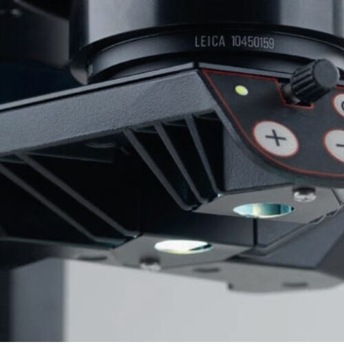
 The unique Leica MCI (Multi Contrast Illumination) systems are ideal for applications that previously required goosenecks. The flat angle of the oblique incident light creates particularly high contrast for precise viewing of even the smallest uneven surfaces and defects such as scratches and dust particles.
 -
 Diffused illumination gives uniform illumination without shadow and glare effects. The Leica LED3000 DI and LED5000 HDI are ideal for documenting samples that are difficult to illuminate using other methods.
 -
 For transmitted light applications. The Leica LED3000 BLI makes it easy to equip a microscope system with transmitted light. The illuminator can be combined with standard baseplates or even used as a standalone instrument.
 -
 Leica LED2000 illumination system was developed for the Leica microscopes of the S series, M series and DMS series. The innovative incident illuminator consists of a 4-point ring illuminator and another arc illuminator for additional contrast.
 -
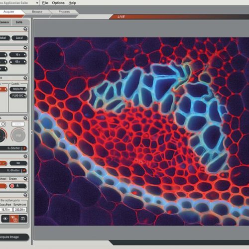 Leica Application Suite X (LAS X) is the one software platform for all Leica microscopes: It integrates confocal, widefield, stereo, super-resolution, and light-sheet instruments from Leica Microsystems.
 -
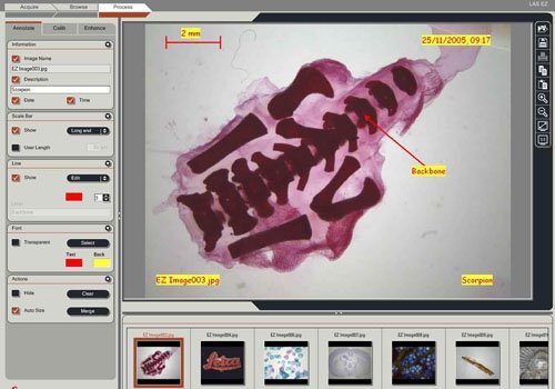 The Leica LAS EZ software, compatible with the Leica EZ4 W/E stereomicroscope, as well as the Leica EC4, Leica ICC50 W/E and Leica IC90 E digital cameras, provides an ideal common, easy-to-use, consistent platform for basic education, industry and life science applications.
 -
 The Leica Keratoscope is a ring illuminator used intraoperatively to qualitatively evaluate the corneal curvature of the eye for astigmatism. With the Leica Keratoscope the surgeon now has a cost-effective, integrated instrument to help assess the shape of the anterior surface of the cornea. This will aid him or her in making limbal relaxing incisions (LRIS) and positioning Toric IOLs.
 -
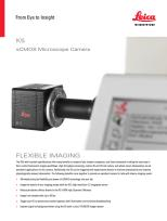 The K5 (grayscale) is ideal for routine fluorescence imaging applications such as immunostaining assays and 3D Cell Culture imaging. Image a wide variety of sample types with the flexibility and power of high 4.2megapixel resolution and fast 40 fps imaging brought to you by the K5.
 -
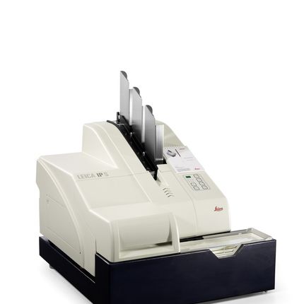 The Leica IP S imprints standard microscope slides, taking only four seconds per imprint when running in serial mode. A patented ink, specially designed for Leica Microsystems, makes imprints resistant to chemical exposure and physical wear. Whether alphanumeric characters, barcodes or logos, the print resolution is always excellent with good legibility. A specific advantage of barcode imprints is the tracking possibility of the probe within the complete histology workflow and additionally the reliable archiving with quick and accurate case identification. An optional external magazine is available to pre-load slides.
 -
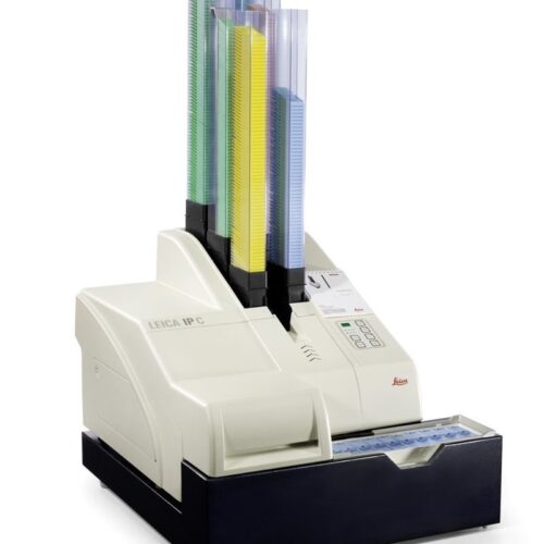 The Leica IP C has been designed for versatile printing of tissue cassettes, including cassettes with lids and two different imprint angles.