
-
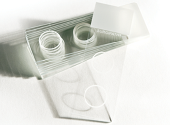 These slides are 3”x 1” White Snowcoat slides with two circles, each having a diameter of 1/2”. These circles match up with the Cytology Funnel, which is to be used for preparation of non-gyn cytology specimens.
 -
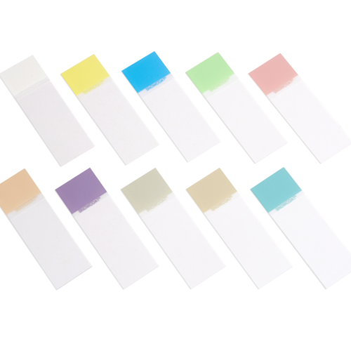 Snowcoat Microscope Slides contain a colored marking area on the surface of the slide to facilitate easy marking/printing and color-coded identification.
 -
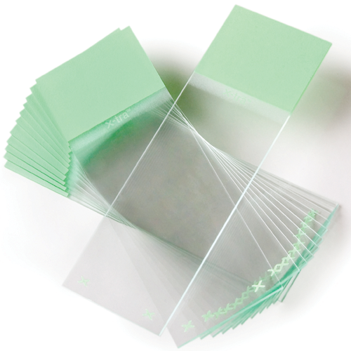 X-tra Slides have a positive charged surface which helps to bond tissue sections and cytology preparations. X‐tra slides are used for demanding H&E procedures that require tissue adhesion. Size: 1” x 3” x .04” (unless otherwise noted)
 -
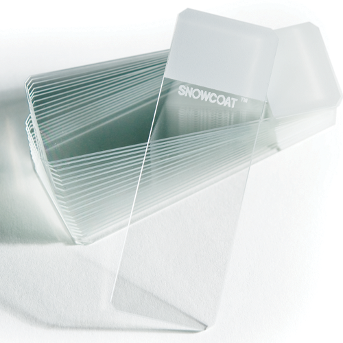 Snowcoat Clipped Corner Slides are clipped at 45° angles to help reduce glass breakage and fragmentation when loaded into slide printers.
 -
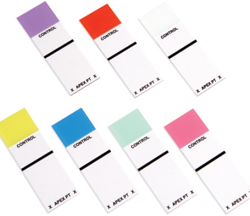 Apex Patient Control Box Slides have clearly designated areas for positive control and patient tissues.
 -
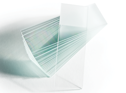 High‐quality sheet glass, manufactured under strict specifications and quality control procedures. Plain and frosted ground versions available.
 -
 The Apex clipped corners are clipped at 45° angles to help reduce glass breakage and fragmentation when loaded into slide printers.
 -
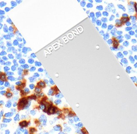 The Apex BOND is an extension of the Apex Adhesive Slides portfolio that is specifically designed and validated for IHC staining on the BOND platform. The eight dots represent the usable areas of the Apex BOND for the different dispense volumes in the BOND processing modules.
 -
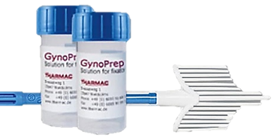 The thin-layer preparation for the gynaecological cytology with the FDA certified Rovers® Cervex-Brush® Combi. Important scientific publication for gynaecologists: Sampling with the Rovers® Cervex-Brush® Combi results in the collection of the same amount of squamous cells, but in a two to threefold harvest of endocervical cells. This leads to the detection of a higher viral load of oncogenic HPV and an increase in the number of detected abnormal smears. Publication in Cytopathology 2006/17 p. 374-381 Improved endocervical sampling and HPV viral load detection by Cervex-Brush® Combi C. E. Depuydt*, I.H. Benoy, E. J. Bailleul**, J. Vandepitte*, A. J. Vereecken* and J. J. Bogers*** *Laboratory for Clinical Pathology (Labo RIATOL), Antwerp, Belgium, **Department of Gynecology, Sint-Vincentius Ziekenhuis, Nortsel, Belgium and ***Laboratory of Cell and Tissue Research, Faculty of Medicine, University of Antwerp, Antwerp, Belgium.
 -
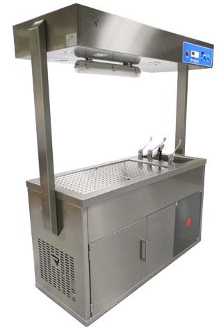 More than 20 years of experience have led us to develop and manufacture a wide variety of dissection and necropsy tables, including the veterinary area. We have a wide range of models (see attached photos).
 -
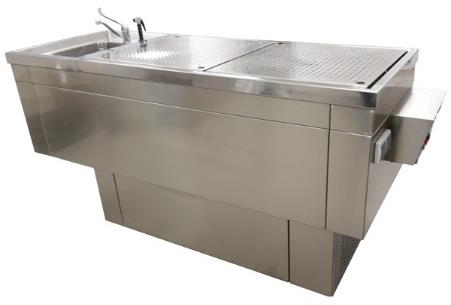 More than 20 years of experience have led us to develop and manufacture a wide variety of dissection and necropsy tables, including the veterinary area. We have a wide range of models (see attached photos).
 -
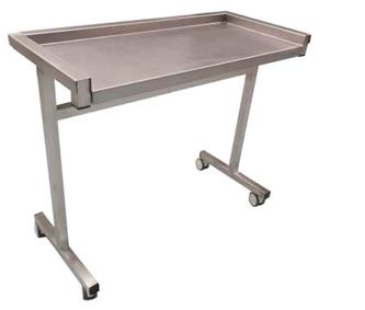 More than 20 years of experience have led us to develop and manufacture a wide variety of dissection and necropsy tables, including the veterinary area. We have a wide range of models (see attached photos).