-
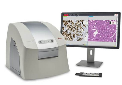 The Aperio LV1 is a cost-effective solution for research users looking to adopt Digital Pathology. The small footprint and compact design makes the Aperio LV1 an ideal desktop system. With minimal IT requirements and rapid implementation, your Aperio LV1 can be installed in minutes.
 -
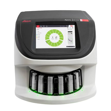 The Aperio GT 450 enables histotechnicians to complete scanning tasks quickly and with confidence leveraging a 32-second scan speed*. Output 81 slides/hr at 40x* delivering high quality images with Leica optics and with an IT architecture that is secure and scalable. From the pathology lab to the IT room, the Aperio GT 450 is designed to scale up digital pathology operations.
 -
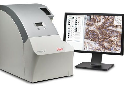 With its compact size, high capacity autoloader and optimal image quality, the Aperio AT2 slide scanner has set new standards in digital pathology. Aperio AT2 consistently delivers precise, whole slide images with an unparalleled, low rescan rate. Slides are available for remote viewing in less than a minute. With an easy-to-use Pathologist's cockpit, coupled with easy integration into laboratory information systems (LIS), the Aperio AT2 provides an ideal platform for research institutions.
 -
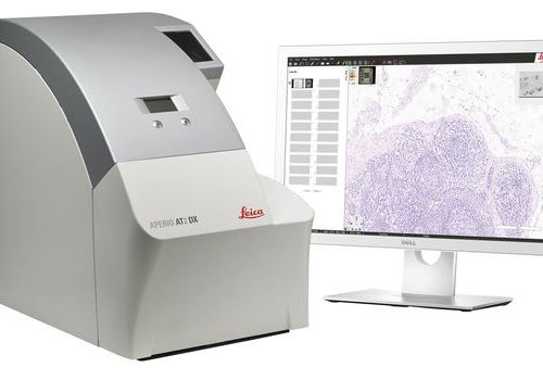 ntroducing the Aperio AT2 DX System, a 400 slide scanner with Aperio ImageScope DX clinical viewing software, and a high-performance viewing workstation including color-calibrated medical grade monitors. The system can be used standalone, or in conjunction with Aperio Path DX case management software, for a streamlined, clinical workflow. Developed with proven technology from Leica Biosystems, the Aperio AT2 DX System enables high volume clinical labs to produce consistent, diagnostic quality images, enabling pathologists to diagnose with confidence.
 -
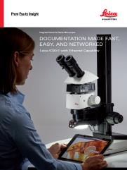 The Leica IC90 E CMOS microscope camera for stereo microscopes was developed for the Leica M series stereo microscopes. It provides pin-sharp images with spot-on color reproduction for professional visualization and documentation of industrial applications.
 -
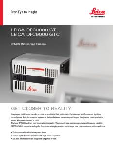 The Leica DFC9000 sCMOS camera will turn your imagination into reality. This monochrome microscope camera with newest scientific CMOS (sCMOS) sensor technology for fluorescence imaging enables you to image your cells under near-native conditions.
 -
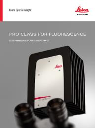 These cooled 2.8 megapixel cameras provide a new paradigm for camera image quality through truly innovative design. The monochrome DFC7000 GT for demanding fluorescence applications, or the color DFC7000 T developed for both brightfield and fluorescence imaging.
 -
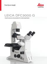 The Leica DFC3000 G is a grayscale USB 3.0 microscope camera for routine fluorescence applications. You will receive crisp images due to its unique passive cooling architecture and its highly sensitive CCD sensor. Correlated pixel double sampling together with a reduction of the ambient temperature of the sensor offers an exceptionally clear and noise -free signal. Its CCD sensor is particularly suitable for low light situations such as fluorescence and will capture even smallest amounts of light.
 -
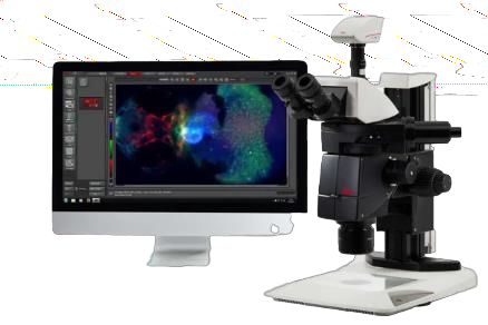
 The K5 (grayscale) is ideal for routine fluorescence imaging applications such as immunostaining assays and 3D Cell Culture imaging. Image a wide variety of sample types with the flexibility and power of high 4.2megapixel resolution and fast 40 fps imaging brought to you by the K5.
 -
 The DMC6200 camera delivers amazing images with intense color detail and contrast - whether at the lowest or highest magnification. Its state-of-the-art CMOS sensor has a 5.86 μm pixel size, 2.3 megapixels sensor resolution, and an astounding dynamic range of 73 dB (4000:1). Achieve an image resolution of up to 20.7 megapixels with cutting-edge pixel shift technology.
 -
 The DMC5400 color CMOS camera offers high frame rate & high-resolution color images even at low magnification. It is optimized to produce fast, high quality images for documentation, evaluation and analysis, for a wide variety of samples and applications in manufacturing and life science research.
 -
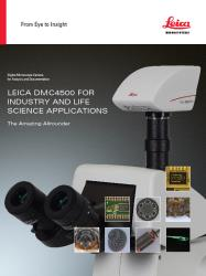 Leica DMC4500 color camera. It has been designed as a versatile, uncomplicated tool that simplifies the imaging process from capture through to processing. It is ideally suited to life science applications such as documenting slides and organisms, pathology or pharmaceutical testing and industrial applications such as quality control and failure analysis.
 -
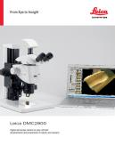 The Leica DMC2900 is a digital USB 3.0 microscope camera with a 3.1 Megapixel CMOS sensor. It is an ideal tool for brightfield microscopic standard applications in research, life science and industry that often require capturing, documenting, and analyzing color images at optimum visibility of microstructures in little time. The Leica DMC2900 is a digital USB 3.0 microscope
 -
 The FLEXACAM C1 can be adapted to your workplace setup - simply connect the camera to your monitor or PC and network. Thanks to the 12 MP CMOS sensor and large dynamic range, fine details and true-to-life colors are a given. adaptable microscope camera solution for a wide variety of samples and applications in industry, life science, forensics, and education .
 -
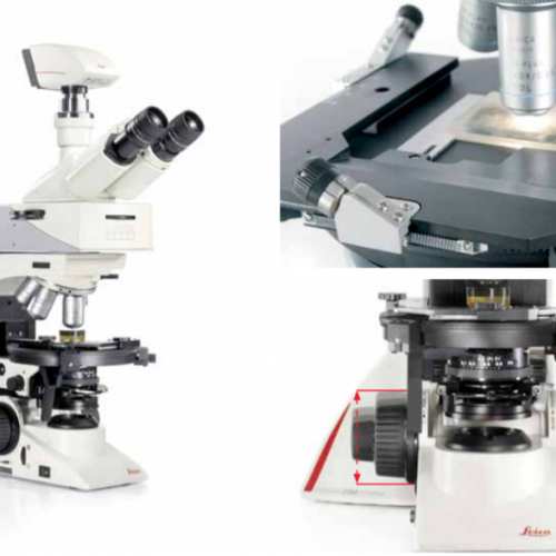
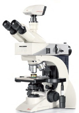 The Leica DM2700 M flexible upright microscope system uses LED illumination for all contrast methods: brightfield (BF), darkfield (DF), differential interference contrast (DIC), qualitative polarization (POL), and fluorescence (FLUO) applications. It also offers built-in oblique illumination, which improves the visualization of surface topography and defects. Optionally, the Leica DM2700 M can also be equipped with a transmitted light axis. The Leica DM2700 M is equipped, e.g. with an N PLAN achromatic objective series with magnifications from 5x to 100x, a field of view of 22 mm, a flattened image field, and large working distances.
 -
The Leica DM2700 M flexible upright microscope system uses LED illumination for all contrast methods: brightfield (BF), darkfield (DF), differential interference contrast (DIC), qualitative polarization (POL), and fluorescence (FLUO) applications. It also offers built-in oblique illumination, which improves the visualization of surface topography and defects. Optionally, the Leica DM2700 M can also be equipped with a transmitted light axis. The Leica DM2700 M is equipped, e.g. with an N PLAN achromatic objective series with magnifications from 5x to 100x, a field of view of 22 mm, a flattened image field, and large working distances.
-
 The Leica DM2700 M flexible upright microscope system uses LED illumination for all contrast methods: brightfield (BF), darkfield (DF), differential interference contrast (DIC), qualitative polarization (POL), and fluorescence (FLUO) applications. It also offers built-in oblique illumination, which improves the visualization of surface topography and defects. Optionally, the Leica DM2700 M can also be equipped with a transmitted light axis. The Leica DM2700 M is equipped, e.g. with an N PLAN achromatic objective series with magnifications from 5x to 100x, a field of view of 22 mm, a flattened image field, and large working distances.
 -
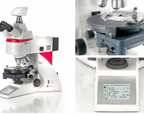
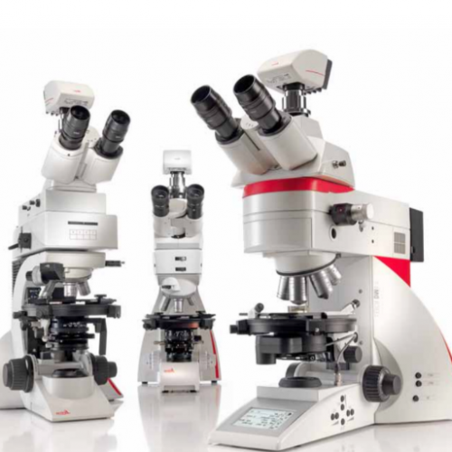 Depending on your application, you can choose from three different systems for polarization microscope. The Leica polarization microscope series is designed for all polarizing examinations: petrography, mineralogy, structure characterization, asbestos analysis, coal analysis (vitrinite reflection), and examination of liquid crystals.
 -
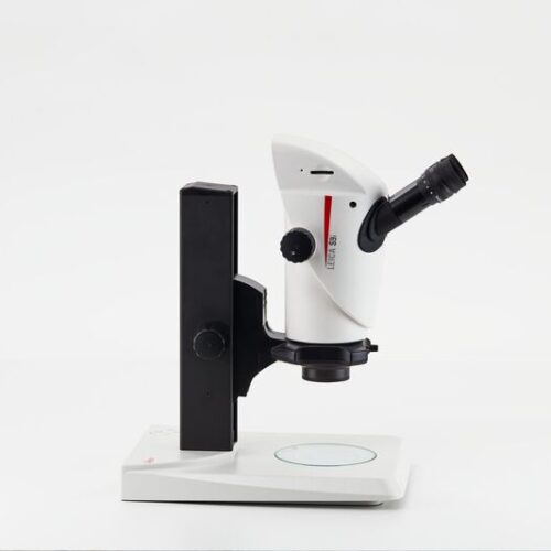 Enjoy working with HD-microscope images live streamed to your PC, HD-monitor, or mobile device. The S9 i stereo microscope has an integrated 10 MP CMOS-camera and can be connected with your facility’s network by Ethernet. This gives you the opportunity to quickly react to queries, get a second opinion, and discuss problems e.g. via tablet with others.
 -
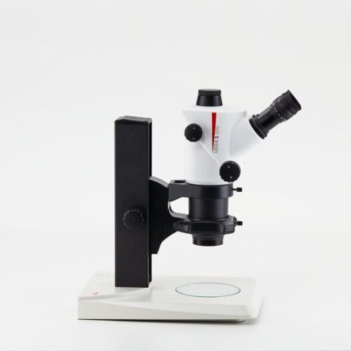 The Greenough stereo microscope, S APO with apochromatic 8:1 zoom and high magnification up to 80x is ideal for quality control, cell sorting, and microinjection applications.
 -
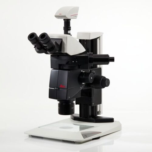
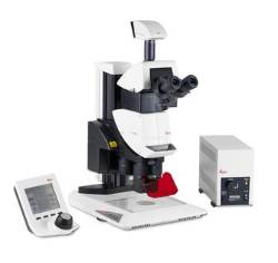 The Leica M205 FA opens a new world of research in fluorescence microscopy, for instance when working in a sterile cabinet.
 -
 Until now, you probably had to switch between two different systems: one for fast screening with a manual zoom that is intuitive to maneuver and a high-end solution to see and capture the faintest signals in an image.
 -
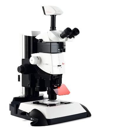 The Leica M165 FC fully apochromatic corrected stereo microscope with 16.5:1 zoom optics resolves structures down to 1.1 micron for detailed fluorescent imaging.