-
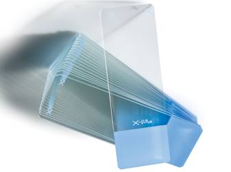
 X-tra Thermal Slides are adhesive slides that are optimized for use with thermal printers. Hydrophobic surface with approximately 90-degree contact angle. Recommended uses include special stains, IHC, ISH, cryosectioning, and cytology.
 -
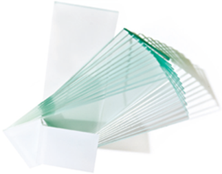
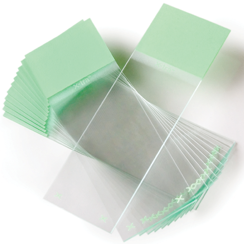 X-tra Slides have a positive charged surface which helps to bond tissue sections and cytology preparations. X‐tra slides are used for demanding H&E procedures that require tissue adhesion. Size: 1” x 3” x .04” (unless otherwise noted)
 -
 X-tra Clipped Corner Slides are clipped at 45° angles to help reduce glass breakage and fragmentation when loaded into slide printers.
 -
 TL3000 Ergo from Leica stands for intuitive manual operation. Effortlessly pass through the different contrast options: brightfield, Rottermann contrast, or one-sided darkfield. Fine-tuning of the contrast is easy, and all with just a single knob.
 -
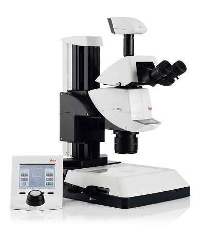
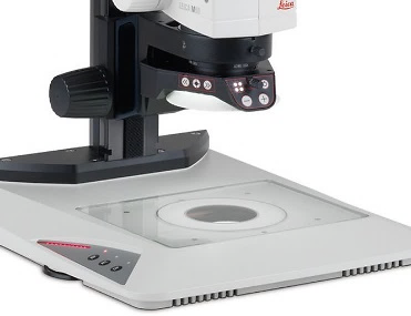 With the TL5000 Ergo you can manage and control all base settings via software for maximum control, whatever your project. Manual push buttons on the base allow cycling through the different contrasts
 -
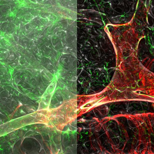 The time has come for imaging systems that allow you to tackle easily biologically relevant 3D models: THUNDER Imagers. To answer important scientific questions, they enable you to obtain a clear view of details, even deep within an intact sample, in real-time without out-of-focus blur.
 -
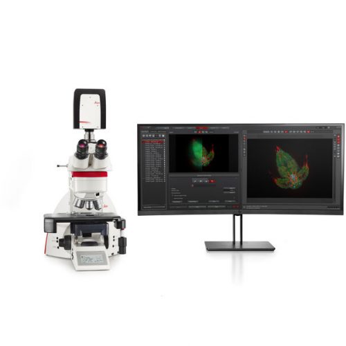 The THUNDER Imager Tissue allows real-time fluorescence imaging of 3D tissue sections typically used in neuroscience and histology research. Acquire rich, detailed images of thick tissues free of haze from out-of-focus blur.
 -
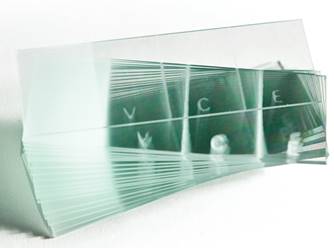 The VCE has two vertical stripes dissecting the slide into three sections allowing vaginal, cervical and endocervical sampling on the same slide. The fully frosted side is completely frosted on one side and is used for body fluid specimens.
 -
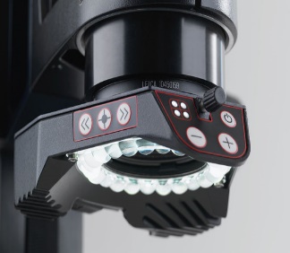 The light is arranged in a ring around the objective and shines down onto the sample. Illuminate from defined segments or uniformly, depending on the properties of the sample
 -
 Snowcoat Thermal Slides are optimized for use with thermal printers.
 -
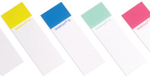
 Snowcoat Pearl slides are cost efficient slides, available in five colors for color-coded identification within the laboratory.
 -
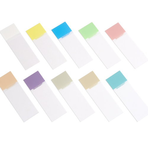 Snowcoat Microscope Slides contain a colored marking area on the surface of the slide to facilitate easy marking/printing and color-coded identification.
 -
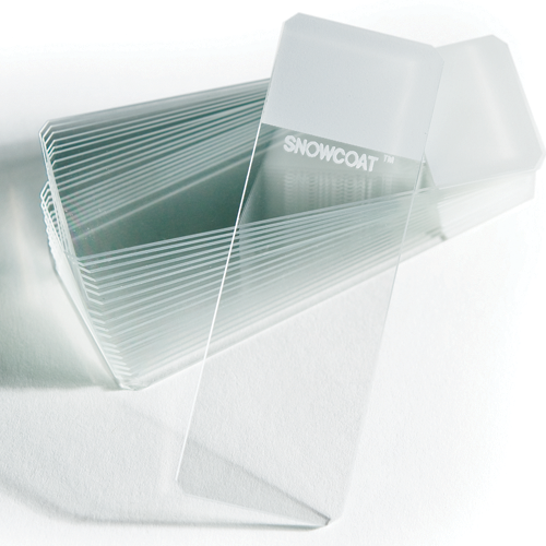 Snowcoat Clipped Corner Slides are clipped at 45° angles to help reduce glass breakage and fragmentation when loaded into slide printers.
 -
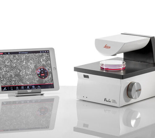 PAULA helps you to speed up your daily cell culture tasks and standardize the results to improve your downstream workflow, offering you essential analysis apps like confluence and transfection efficiency check.
 -
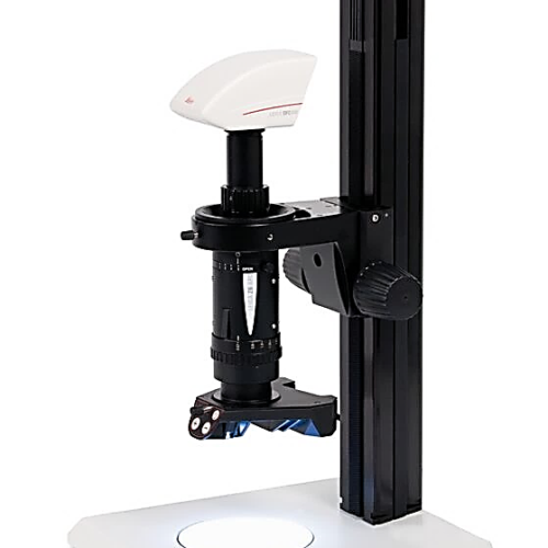
 The purpose of the Leica motor-focus system is to provide motor-driven coarse and fine focusing by means of a manual control, a footswitch, or through a computer. Five exact focus settings can be stored.
 -
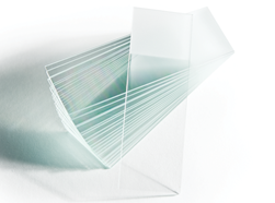 High‐quality sheet glass, manufactured under strict specifications and quality control procedures. Plain and frosted ground versions available.
 -
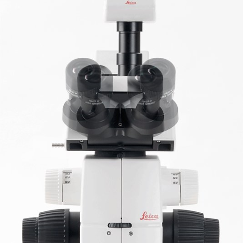
 Manual 6:1 Macroscope for detailed Documentation, Measurement and Evaluation. The Leica Z6 APO is a fully apochromatic zoom system with excellent light transmission for high contrast, high-resolution, detailed analysis. The single beam path provides 2D images and ensures parallax-free imaging. the Leica Z6 APO with 6.3:1 zoom offers the highest numerical aperture: 0.117 nA (351 Lp/mm resolution) with the 1× plan-apochromatic objective
 -
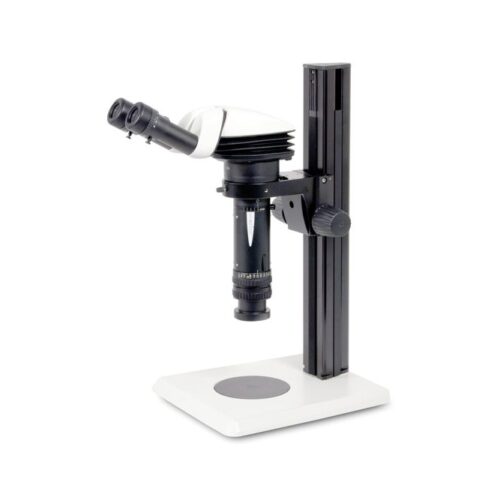 The Leica Z6 APO is a fully apochromatic zoom system with excellent light transmission for high contrast, high-resolution, detailed analysis. The single beam path provides 2D images and ensures parallax-free imaging.
 -
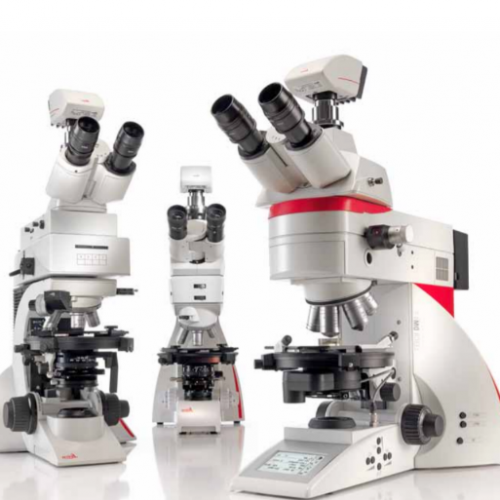 Depending on your application, you can choose from three different systems for polarization microscope. The Leica polarization microscope series is designed for all polarizing examinations: petrography, mineralogy, structure characterization, asbestos analysis, coal analysis (vitrinite reflection), and examination of liquid crystals.
 -
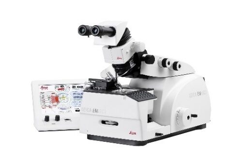 The Leica EM UC7 prepares excellent quality semi- and ultra-thin sections, as well as the perfectly smooth surfaces required for LM, TEM, SEM, and AFM examination. The precision mechanics, ergonomic design, and intuitive layout of the touchscreen control unit make the Leica EM UC7 ideal for the highest quality specimen preparation
 -
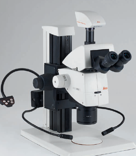
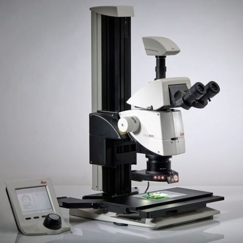
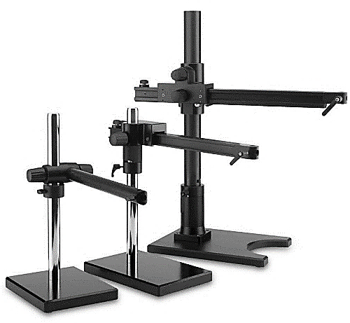 Leica Swingarm Stands (boom stands) are ideal to handle large samples. The high modularity allows to customize these stands depending on sample size and weight of the configuration. Focus arms and drives provide multiple mounting options and more safety features.
 -
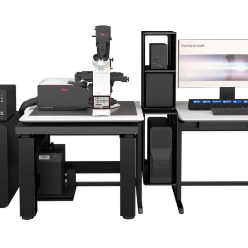 STELLARIS has been designed from the ground up to give you the power to see more. The synergy between the new Power HyD detectors family, the completely optimized beam path, and the new generation White Light Lasers (WLL) empowers you to obtain more accurate and reliable data to test your hypothesis with precision.
 -
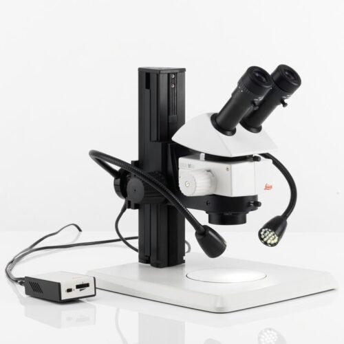
 The Leica LED3000 SLI and Leica LED5000 SLI each feature two bright LED spotlights, which can be individually adjusted to the sample with two goosenecks. The control element is mounted on a separate gooseneck and can be placed in any position.
 -
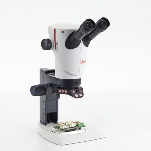
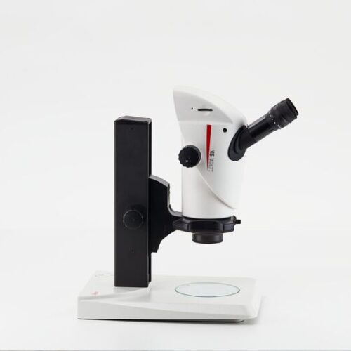 Enjoy working with HD-microscope images live streamed to your PC, HD-monitor, or mobile device. The S9 i stereo microscope has an integrated 10 MP CMOS-camera and can be connected with your facility’s network by Ethernet. This gives you the opportunity to quickly react to queries, get a second opinion, and discuss problems e.g. via tablet with others.
 -
 The S9 D gives you the flexibility to add a camera to the microscope and turn it into a digital microscope solution. The built in 50:50 split of the beam path allows observation through the eyepieces and digital imaging at the same time.
 -
 The Greenough stereo microscope, S APO with apochromatic 8:1 zoom and high magnification up to 80x is ideal for quality control, cell sorting, and microinjection applications.
 -
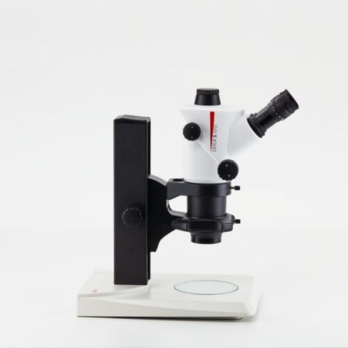
 For analog microscope inspections the S9 E is an indispensable asset. It provides a cost-effective solution with fast return on investment.
 -
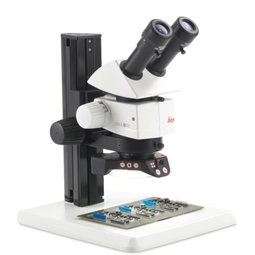
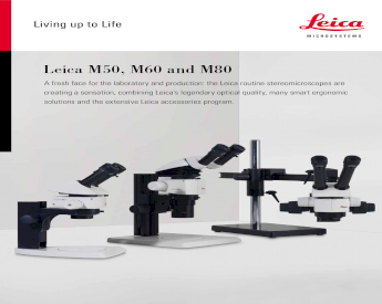 The M50, M60, and M80 allow operators to see a large sample overview, work comfortably under the microscope, and capture images of important details easily.
 -
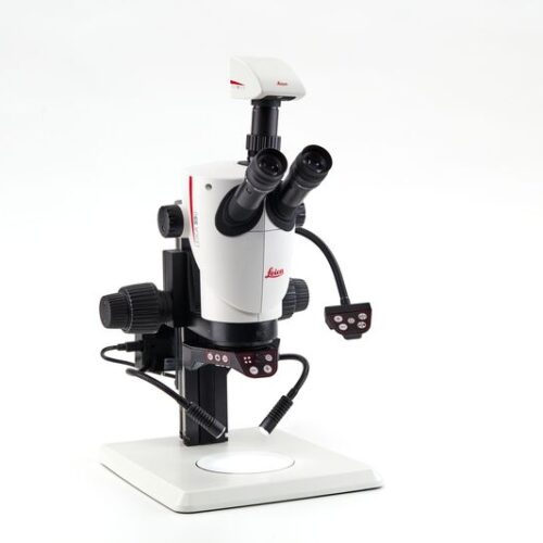
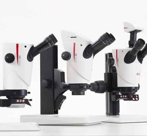 Continuously improving production, keeping defect rates low, and fulfilling customer requests to stay competitive can be very challenging. Leica has developed the S9 stereo microscope series to help you cope with these challenges. With this new generation of Greenough stereo microscopes operators will be able to reveal details faster as they spend less time having to adjust the microscope.
 -
 With a Leica M-series stereo microscope you can choose from a comprehensive range of accessories to adapt your microscope to your application Our goal: high-quality images that allow measurements and can be accurately repeated with minimal effort. The Leica coded stereo microscopes help you to pave the way to correct results.
 -
 M80 with 8:1-zoom range, with a continuous zoom from 7.5x – 60x including engageable click stops – expanding your insights and reliability. Optimized for higher magnification to see smaller details in perfect contrast. The large working distance and brilliant imaging power show the finest details of your specimens without losing the field of view over large specimens
 -
 M60 with 6:1-zoom range, with a continuous zoom from 6.3x – 40x including engageable click stops - making repetitive inspection and measurement at a specific zoom position reliable. This system is optimized for a larger field of view to see more at a glance.
 -
 with 5:1-zoom, featuring a step-zoom with five fixed zoom steps, at 6.3x, 10x, 16x, 25x, and 40x– allowing 100% reproducible results, this ensures that the results remain comparable at all times without great effort. The short optics carrier enables high light throughput and provides a large working distance, ideal in animal surgery and similar applications.
 -
 Imagine not having to choose between high resolution or better depth of field, but to have both! The revolutionary FusionOptics™ technology makes it possible and provides you an ideal 3D image to see even the smallest details.
 -
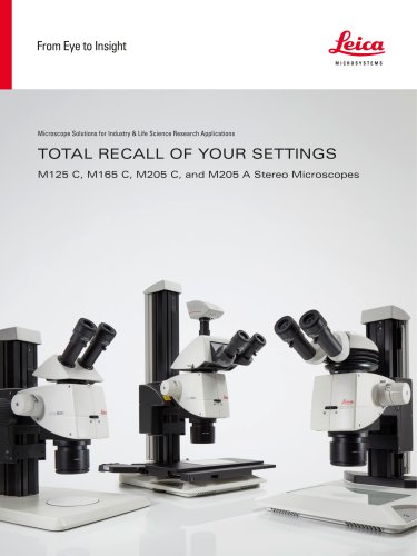
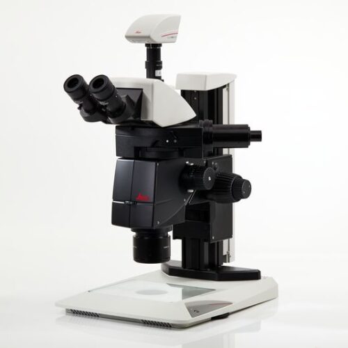
 The Leica M205 C, the world’s first stereomicroscope to offer 20.5:1 zoom, uses unique FusionOptics™ technology to provide exceptional image detail and impressive 3D depth perception. Its rigid, sturdy mechanical structure, revolutionary FusionOptics™, and completely integrated multi-contrast illumination, make the M205 C an ideal instrument for questioned document examiners seeking a high-performance microscope capable of revealing fine details.
 -
 Until now, you probably had to switch between two different systems: one for fast screening with a manual zoom that is intuitive to maneuver and a high-end solution to see and capture the faintest signals in an image.