-
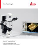 The Leica DMC2900 is a digital USB 3.0 microscope camera with a 3.1 Megapixel CMOS sensor. It is an ideal tool for brightfield microscopic standard applications in research, life science and industry that often require capturing, documenting, and analyzing color images at optimum visibility of microstructures in little time. The Leica DMC2900 is a digital USB 3.0 microscope
 -
 The FLEXACAM C1 can be adapted to your workplace setup - simply connect the camera to your monitor or PC and network. Thanks to the 12 MP CMOS sensor and large dynamic range, fine details and true-to-life colors are a given. adaptable microscope camera solution for a wide variety of samples and applications in industry, life science, forensics, and education .
 -
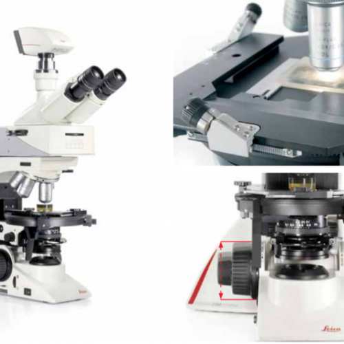
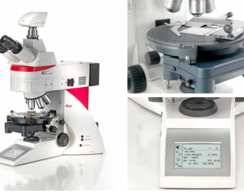
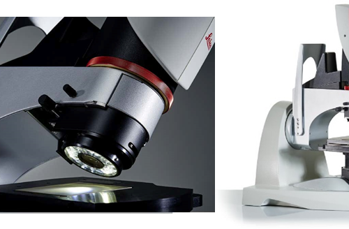
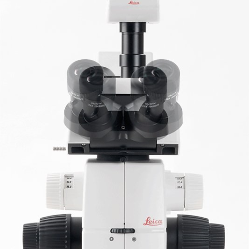
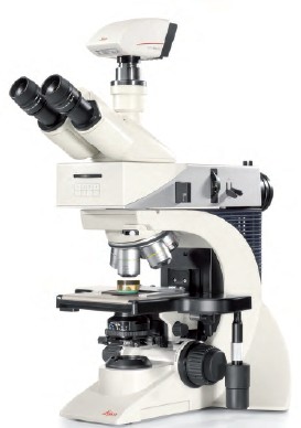 The Leica DM2700 M flexible upright microscope system uses LED illumination for all contrast methods: brightfield (BF), darkfield (DF), differential interference contrast (DIC), qualitative polarization (POL), and fluorescence (FLUO) applications. It also offers built-in oblique illumination, which improves the visualization of surface topography and defects. Optionally, the Leica DM2700 M can also be equipped with a transmitted light axis. The Leica DM2700 M is equipped, e.g. with an N PLAN achromatic objective series with magnifications from 5x to 100x, a field of view of 22 mm, a flattened image field, and large working distances.
 -
The Leica DM2700 M flexible upright microscope system uses LED illumination for all contrast methods: brightfield (BF), darkfield (DF), differential interference contrast (DIC), qualitative polarization (POL), and fluorescence (FLUO) applications. It also offers built-in oblique illumination, which improves the visualization of surface topography and defects. Optionally, the Leica DM2700 M can also be equipped with a transmitted light axis. The Leica DM2700 M is equipped, e.g. with an N PLAN achromatic objective series with magnifications from 5x to 100x, a field of view of 22 mm, a flattened image field, and large working distances.
-
 The Leica DM2700 M flexible upright microscope system uses LED illumination for all contrast methods: brightfield (BF), darkfield (DF), differential interference contrast (DIC), qualitative polarization (POL), and fluorescence (FLUO) applications. It also offers built-in oblique illumination, which improves the visualization of surface topography and defects. Optionally, the Leica DM2700 M can also be equipped with a transmitted light axis. The Leica DM2700 M is equipped, e.g. with an N PLAN achromatic objective series with magnifications from 5x to 100x, a field of view of 22 mm, a flattened image field, and large working distances.
 -
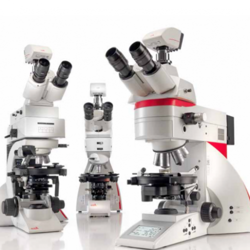 Depending on your application, you can choose from three different systems for polarization microscope. The Leica polarization microscope series is designed for all polarizing examinations: petrography, mineralogy, structure characterization, asbestos analysis, coal analysis (vitrinite reflection), and examination of liquid crystals.
 -
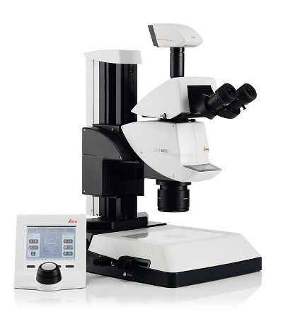
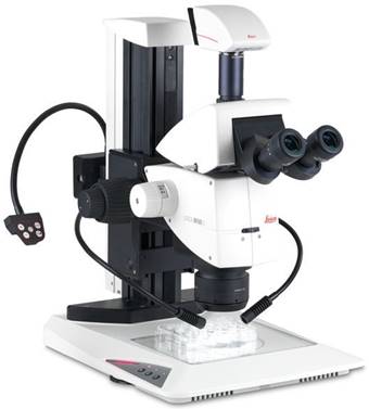
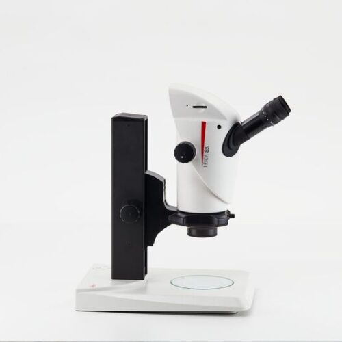 Enjoy working with HD-microscope images live streamed to your PC, HD-monitor, or mobile device. The S9 i stereo microscope has an integrated 10 MP CMOS-camera and can be connected with your facility’s network by Ethernet. This gives you the opportunity to quickly react to queries, get a second opinion, and discuss problems e.g. via tablet with others.
 -
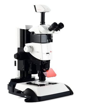
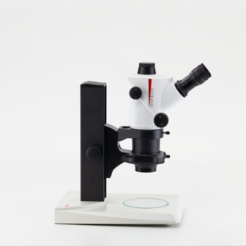 The Greenough stereo microscope, S APO with apochromatic 8:1 zoom and high magnification up to 80x is ideal for quality control, cell sorting, and microinjection applications.
 -
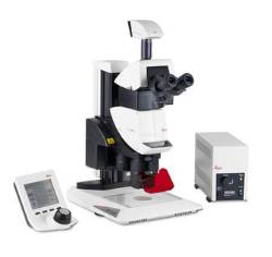 The Leica M205 FA opens a new world of research in fluorescence microscopy, for instance when working in a sterile cabinet.
 -
 Until now, you probably had to switch between two different systems: one for fast screening with a manual zoom that is intuitive to maneuver and a high-end solution to see and capture the faintest signals in an image.
 -
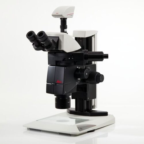
 The Leica M165 FC fully apochromatic corrected stereo microscope with 16.5:1 zoom optics resolves structures down to 1.1 micron for detailed fluorescent imaging.
 -
 Imagine not having to choose between high resolution or better depth of field, but to have both! The revolutionary FusionOptics™ technology makes it possible and provides you an ideal 3D image to see even the smallest details.
 -
 With the DVM6, you can get from the big picture to smallest details in an instant. You can seamlessly carry on working even if changing objective is required, as the sample always stays in focus and no pre-adjustments are needed.
 -
 The intuitive A60 F and A60 S stereo microscopes fulfill what you need – high sample throughput, optimum visibility of product details and components, and easy processing of subassemblies.
 -
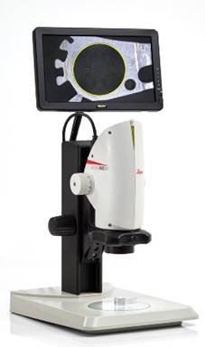 Digital microscope system for digital inspection, observation and measurement. From tiniest detail to an overview, the optics ensures magnification of up to 300x. The built-in HDMI microscope camera provides full high definition live images of up to 30fps and a resolution of 5Mpixels.
 -
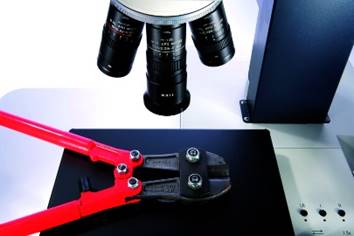
 With a Leica M-series stereo microscope you can choose from a comprehensive range of accessories to adapt your microscope to your application, whether it is in materials testing, life science research or forensic application
 -
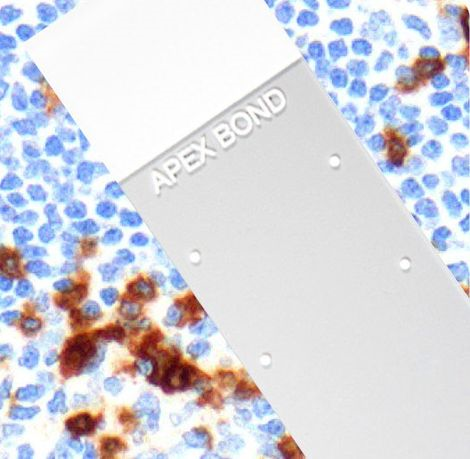 The Apex BOND is an extension of the Apex Adhesive Slides portfolio that is specifically designed and validated for IHC staining on the BOND platform. The eight dots represent the usable areas of the Apex BOND for the different dispense volumes in the BOND processing modules.
 -
 The Apex clipped corners are clipped at 45° angles to help reduce glass breakage and fragmentation when loaded into slide printers.
 -
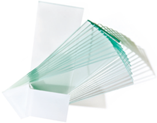
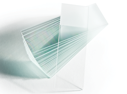 High‐quality sheet glass, manufactured under strict specifications and quality control procedures. Plain and frosted ground versions available.
 -
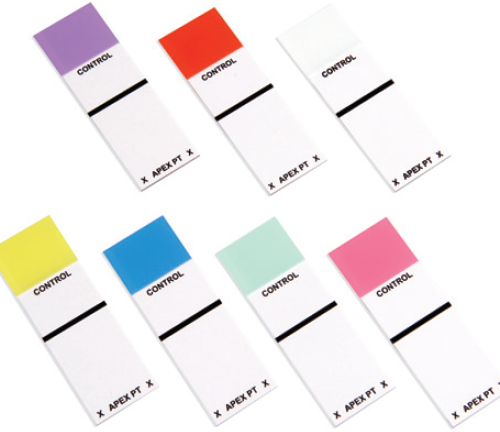 Apex Patient Control Box Slides have clearly designated areas for positive control and patient tissues.
 -
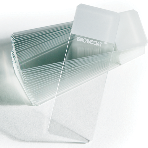 Snowcoat Clipped Corner Slides are clipped at 45° angles to help reduce glass breakage and fragmentation when loaded into slide printers.
 -
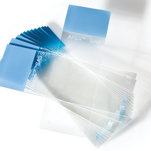 The Apex Adhesive Slides portfolio consists of microscope slides made of white glass and coated with a proprietary adhesive coating for a positive charge, to promote tissue adhesion and to allow the slide to be more hydrophilic.
 -
 X-tra Thermal Slides are adhesive slides that are optimized for use with thermal printers. Hydrophobic surface with approximately 90-degree contact angle. Recommended uses include special stains, IHC, ISH, cryosectioning, and cytology.
 -
 Snowcoat Thermal Slides are optimized for use with thermal printers.
 -
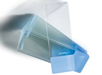
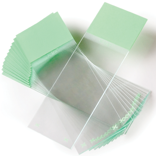 X-tra Slides have a positive charged surface which helps to bond tissue sections and cytology preparations. X‐tra slides are used for demanding H&E procedures that require tissue adhesion. Size: 1” x 3” x .04” (unless otherwise noted)
 -
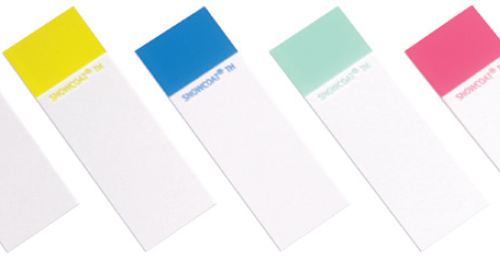
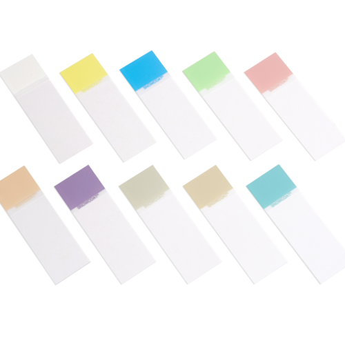 Snowcoat Microscope Slides contain a colored marking area on the surface of the slide to facilitate easy marking/printing and color-coded identification.
 -
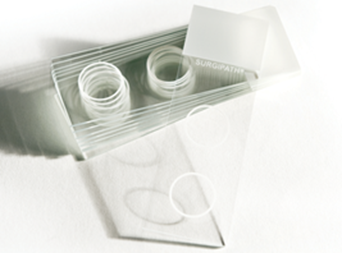 These slides are 3”x 1” White Snowcoat slides with two circles, each having a diameter of 1/2”. These circles match up with the Cytology Funnel, which is to be used for preparation of non-gyn cytology specimens.
 -
 Snowcoat Pearl slides are cost efficient slides, available in five colors for color-coded identification within the laboratory.
 -
 X-tra Clipped Corner Slides are clipped at 45° angles to help reduce glass breakage and fragmentation when loaded into slide printers.
 -
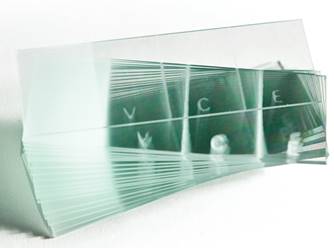 The VCE has two vertical stripes dissecting the slide into three sections allowing vaginal, cervical and endocervical sampling on the same slide. The fully frosted side is completely frosted on one side and is used for body fluid specimens.
 -
 Beveled Edge Slides feature beveled edges and corners that make for easier handling. The 25 mm x 75mm slides are washed and carefully inspected to ensure quality.
 -
 These absolute precision scales have a large 13 inches (330 mm) double dial with chrome-plated housing. The scales have large numerals and graduations for easy reading. The removable pan is constructed of stainless Steel and features drain holes
 -
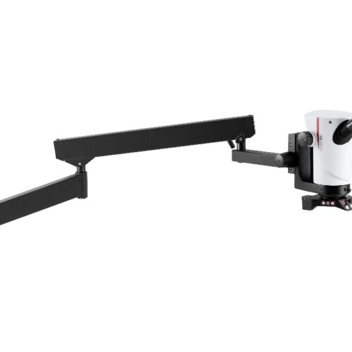 The Leica Flexarm stand can rotate 360° and has a radius of up to one meter, making it highly mobile. This allows for convenient viewing of large samples, which should not or cannot be moved. This is ideal for inspection, assembly, and quality control in electronics manufacturing, in dental laboratories and many other applications.
 -
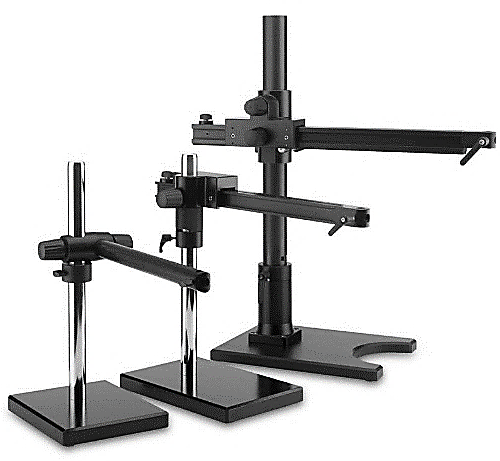 Leica Swingarm Stands (boom stands) are ideal to handle large samples. The high modularity allows to customize these stands depending on sample size and weight of the configuration. Focus arms and drives provide multiple mounting options and more safety features.
 -
 The purpose of the Leica motor-focus system is to provide motor-driven coarse and fine focusing by means of a manual control, a footswitch, or through a computer. Five exact focus settings can be stored.