-
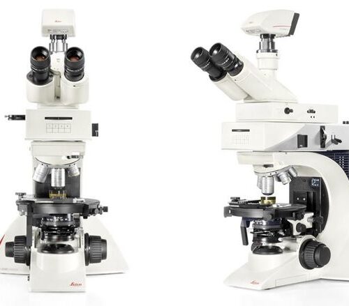
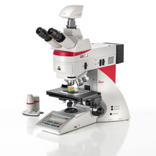
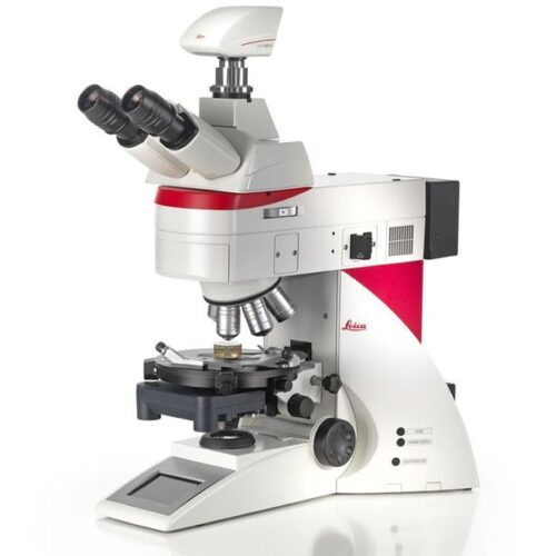
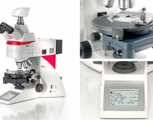
 The Leica DM2700 M flexible upright microscope system uses LED illumination for all contrast methods: brightfield (BF), darkfield (DF), differential interference contrast (DIC), qualitative polarization (POL), and fluorescence (FLUO) applications. It also offers built-in oblique illumination, which improves the visualization of surface topography and defects. Optionally, the Leica DM2700 M can also be equipped with a transmitted light axis. The Leica DM2700 M is equipped, e.g. with an N PLAN achromatic objective series with magnifications from 5x to 100x, a field of view of 22 mm, a flattened image field, and large working distances.
 -
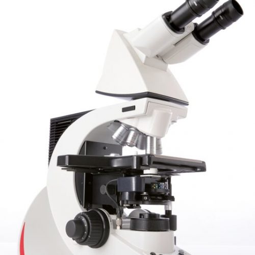
 The Leica DM2700 P offers a level of reproducibility that is one-of-a-kind in its class, the built-in focus stop protects your samples and the front lens of the objective. For samples of equal height, the focus stop makes the focusing plane easier to reconstruct so you can concentrate entirely on your application. Color-coded lenses match the color-coded field and aperture diaphragm adjustment (CDA), to ensure that the illumination conditions are always matched to the objective. Constant color temperature by advanced LED technology.
 -
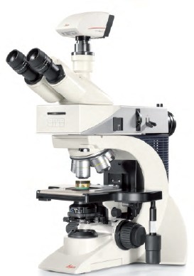 The Leica DM2700 M flexible upright microscope system uses LED illumination for all contrast methods: brightfield (BF), darkfield (DF), differential interference contrast (DIC), qualitative polarization (POL), and fluorescence (FLUO) applications. It also offers built-in oblique illumination, which improves the visualization of surface topography and defects. Optionally, the Leica DM2700 M can also be equipped with a transmitted light axis. The Leica DM2700 M is equipped, e.g. with an N PLAN achromatic objective series with magnifications from 5x to 100x, a field of view of 22 mm, a flattened image field, and large working distances.
 -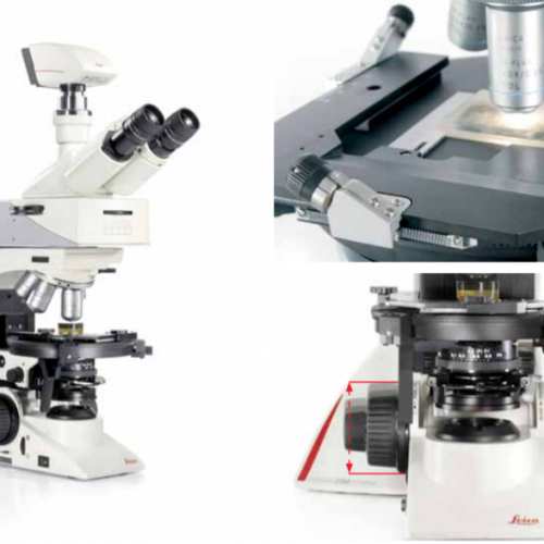
 The Leica DM3000 microscopes for pathology, cytology, haematology and many other applications feature a motorized nosepiece, condenser head, automated light intensity adjustment, and optional foot pedal. These intuitive microscopes improve workflows significantly. For special diagnostics requirements, the microscope is certified for in-vitro-diagnostics (IVD) like in-vitro-fertilization (IVF).
 -
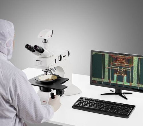 System for Microelectronics and Semiconductor. With a large field of view, the DM3 XL inspection system allows your team to identify defects faster and increase your yield rate. Make use of the 30% increased field of view of the unique macro objective. The DM3 XL uses LED illumination for all contrast methods. LED illumination provides a constant color temperature and offers real color imaging at all intensity levels.
 -
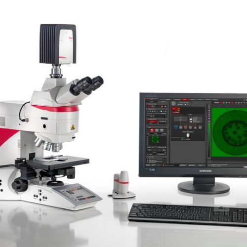
 Increase your work efficiency with the Leica DM4 B or Leica DM6 B upright digital research microscopes! Both are ideally suited to making your work life easier in biomedical research and clinical labs alike. You can configure the microscope that fits your need and budget with the Leica DM4 B and Leica DM6 B. Optimize your system to your application with different contrast methods and automated functions.
 -
 Fluorescence is one of the most commonly used physical phenomena in biological and analytical microscopy, mainly because of its high sensitivity and high specificity. Fluorescence is a form of luminescence.
 -
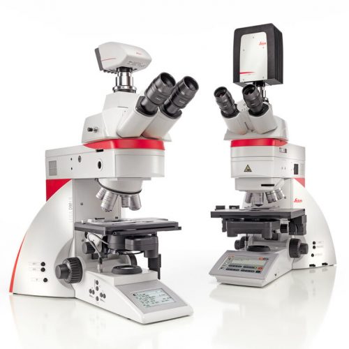
 Leica DM4 M and DM6 M digital microscopes for materials science and quality control offer truly reproducible microscopy, incredible optics and high-quality images. Store and recall your imaging conditions with a touch of a button. High quality microscope images make challenging inspection, measurement, and analysis tasks easy. Use the Leica DM4 M for manual routine inspection or the Leica DM6 M for fully automated materials analysis.
 -
 Fully coded and semi-automated. The Leica DM4 P automatically detects which contrast method and objective are being used. This provides valuable consistency and reproducibility for your research. Manual diaphragm setting is no longer required, either in the transmitted light or incident light method. Light intensity automatically adjusts to the objective. Image brightness remains constant when switching objectives, which eliminates glare.
 -
 The Leica DM2700 M flexible upright microscope system uses LED illumination for all contrast methods: brightfield (BF), darkfield (DF), differential interference contrast (DIC), qualitative polarization (POL), and fluorescence (FLUO) applications. It also offers built-in oblique illumination, which improves the visualization of surface topography and defects. Optionally, the Leica DM2700 M can also be equipped with a transmitted light axis. The Leica DM2700 M is equipped, e.g. with an N PLAN achromatic objective series with magnifications from 5x to 100x, a field of view of 22 mm, a flattened image field, and large working distances.
 -
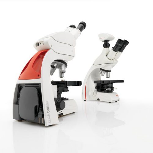 The Leica DM500 microscope with “plug and play” capability is the ideal tool to make teaching entry-level college and university Life Science courses easy and fun for the instructor and the student.