-
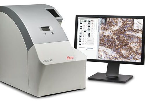 With its compact size, high capacity autoloader and optimal image quality, the Aperio AT2 slide scanner has set new standards in digital pathology. Aperio AT2 consistently delivers precise, whole slide images with an unparalleled, low rescan rate. Slides are available for remote viewing in less than a minute. With an easy-to-use Pathologist's cockpit, coupled with easy integration into laboratory information systems (LIS), the Aperio AT2 provides an ideal platform for research institutions.
 -
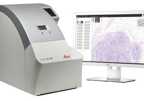 ntroducing the Aperio AT2 DX System, a 400 slide scanner with Aperio ImageScope DX clinical viewing software, and a high-performance viewing workstation including color-calibrated medical grade monitors. The system can be used standalone, or in conjunction with Aperio Path DX case management software, for a streamlined, clinical workflow. Developed with proven technology from Leica Biosystems, the Aperio AT2 DX System enables high volume clinical labs to produce consistent, diagnostic quality images, enabling pathologists to diagnose with confidence.
 -
 The Leica IC90 E CMOS microscope camera for stereo microscopes was developed for the Leica M series stereo microscopes. It provides pin-sharp images with spot-on color reproduction for professional visualization and documentation of industrial applications.
 -
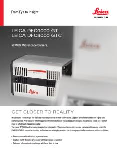 The Leica DFC9000 sCMOS camera will turn your imagination into reality. This monochrome microscope camera with newest scientific CMOS (sCMOS) sensor technology for fluorescence imaging enables you to image your cells under near-native conditions.
 -
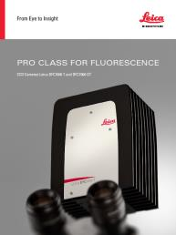 These cooled 2.8 megapixel cameras provide a new paradigm for camera image quality through truly innovative design. The monochrome DFC7000 GT for demanding fluorescence applications, or the color DFC7000 T developed for both brightfield and fluorescence imaging.
 -
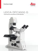 The Leica DFC3000 G is a grayscale USB 3.0 microscope camera for routine fluorescence applications. You will receive crisp images due to its unique passive cooling architecture and its highly sensitive CCD sensor. Correlated pixel double sampling together with a reduction of the ambient temperature of the sensor offers an exceptionally clear and noise -free signal. Its CCD sensor is particularly suitable for low light situations such as fluorescence and will capture even smallest amounts of light.
 -
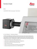 The K5 (grayscale) is ideal for routine fluorescence imaging applications such as immunostaining assays and 3D Cell Culture imaging. Image a wide variety of sample types with the flexibility and power of high 4.2megapixel resolution and fast 40 fps imaging brought to you by the K5.
 -
 The DMC6200 camera delivers amazing images with intense color detail and contrast - whether at the lowest or highest magnification. Its state-of-the-art CMOS sensor has a 5.86 μm pixel size, 2.3 megapixels sensor resolution, and an astounding dynamic range of 73 dB (4000:1). Achieve an image resolution of up to 20.7 megapixels with cutting-edge pixel shift technology.
 -
 The DMC5400 color CMOS camera offers high frame rate & high-resolution color images even at low magnification. It is optimized to produce fast, high quality images for documentation, evaluation and analysis, for a wide variety of samples and applications in manufacturing and life science research.
 -
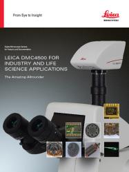 Leica DMC4500 color camera. It has been designed as a versatile, uncomplicated tool that simplifies the imaging process from capture through to processing. It is ideally suited to life science applications such as documenting slides and organisms, pathology or pharmaceutical testing and industrial applications such as quality control and failure analysis.
 -
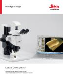 The Leica DMC2900 is a digital USB 3.0 microscope camera with a 3.1 Megapixel CMOS sensor. It is an ideal tool for brightfield microscopic standard applications in research, life science and industry that often require capturing, documenting, and analyzing color images at optimum visibility of microstructures in little time. The Leica DMC2900 is a digital USB 3.0 microscope
 -
 The FLEXACAM C1 can be adapted to your workplace setup - simply connect the camera to your monitor or PC and network. Thanks to the 12 MP CMOS sensor and large dynamic range, fine details and true-to-life colors are a given. adaptable microscope camera solution for a wide variety of samples and applications in industry, life science, forensics, and education .
 -
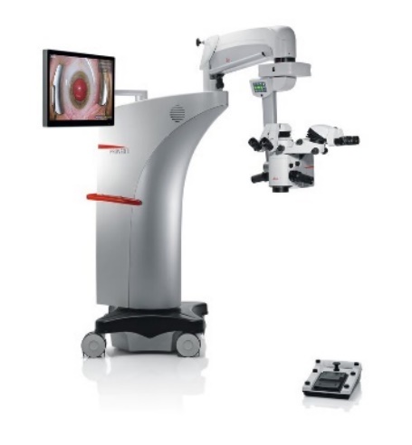 The Proveo 8 ophthalmic microscope provides the exact image you need at each moment of your procedure. Like a precision timepiece every element of the Proveo 8 microscope interconnects and works in perfect synchrony to optimize your view.
 -
 The Leica M844 F40 belongs to the premium class of ophthalmic surgical microscopes and offers optimal solutions particularly for posterior and high-end anterior segment surgery.
 -
 The Leica M822 surgical microscope, with enhanced Red Reflex, meets the highest professional requirements of ophthalmic surgeons to perform cataract surgery more precisely and efficiently.
 -
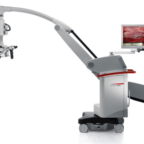
 The Leica OH4 surgical microscope provides superior reach, height, and clearance. The Leica OH4 floor stands that moves easily and when the brakes are released, the microscope stabilizes in only 1.5 seconds and remains vibration-free. By integrating new glass, glass coatings, and design parameters, high-resolution APO OptiChrome technology delivers an expanded working distance and greater depth of focus. In combination with the innovative light management systems BrightCare and AutoIris, you can operate and safe light levels and still see more than ever before. The M525 OH4 also offers FL800 and FL400 fluorescence for intraoperative visualization of blood flow and tumour tissue.
 -
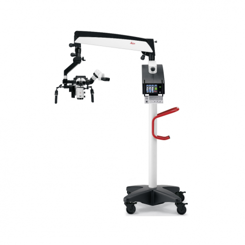
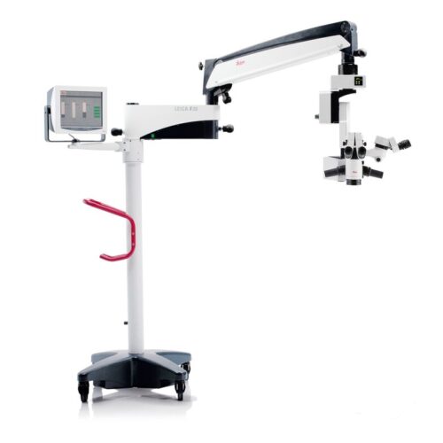
Compact surgical microscope Smooth, fine movements, Xenon illumination systems, Motorized MultiFoc lens
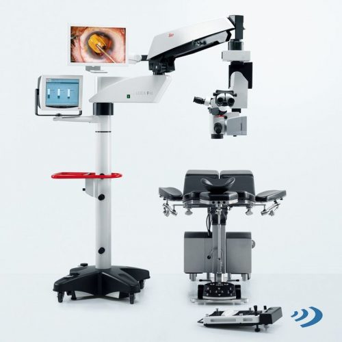
The Leica M525 F20 surgical microscope combines brilliant optical quality with superior maneuverability. The crisp, sharp images and large depth of field allow the surgeon to see precise details. The working-distance-based illumination provides enough light for deep operative sites while supporting patient safety. Designed as an otolaryngology microscope, the Leica M525 F20 is also ideal for spine, dental, hand, and plastic/reconstructive procedures. Ease of movement -
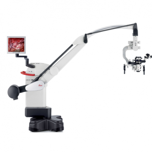
Precision Surgical Microscope For Neurosurgery, Spine & Plastic Reconstructive Surgery
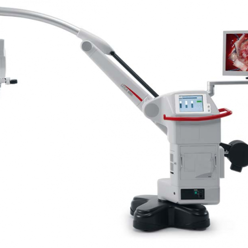
Precision comes standard in our M530 OHX, helping you achieve the highest standards of precision in your surgery. -
Neurosurgery Microscope
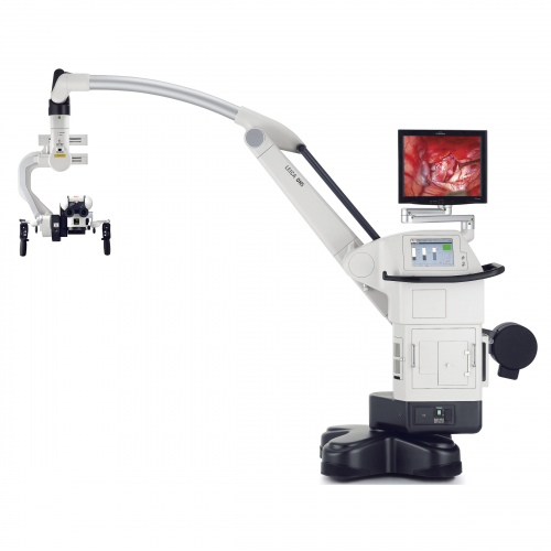
The Leica M530 OH6 takes image quality to a new level: FusionOptics, the groundbreaking technology by Leica Microsystems, unites an enhanced depth of field with high resolution to create an optimal view. FusionOptics also helps you to streamline your work: A larger area in full focus means that there is less need to refocus the microscope. FusionOptic let you stay focused – in every sense of the word. -
Premium Surgical Microscope for Microsurgery
The Leica M720 OH5 is the premium operating microscope for neurosurgery, otolaryngology and reconstructive microsurgery. With this operating microscope, surgeons will benefit from greater visibility in deep cavities or lesions and improved ergonomics and comfort. The Leica M720 OH5 surgical microscope also offers full HD 3D and 2D visualization and recording, so that the entire surgical team as well as students will be able to observe and learn more easily. The robust OH5 floor stand, designed by Mitaka, features long reach and electromagnetic brakes for lightweight positioning and fast stabilization. -
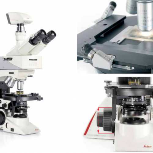
 The Leica DM2700 M flexible upright microscope system uses LED illumination for all contrast methods: brightfield (BF), darkfield (DF), differential interference contrast (DIC), qualitative polarization (POL), and fluorescence (FLUO) applications. It also offers built-in oblique illumination, which improves the visualization of surface topography and defects. Optionally, the Leica DM2700 M can also be equipped with a transmitted light axis. The Leica DM2700 M is equipped, e.g. with an N PLAN achromatic objective series with magnifications from 5x to 100x, a field of view of 22 mm, a flattened image field, and large working distances.
 -
The Leica DM2700 M flexible upright microscope system uses LED illumination for all contrast methods: brightfield (BF), darkfield (DF), differential interference contrast (DIC), qualitative polarization (POL), and fluorescence (FLUO) applications. It also offers built-in oblique illumination, which improves the visualization of surface topography and defects. Optionally, the Leica DM2700 M can also be equipped with a transmitted light axis. The Leica DM2700 M is equipped, e.g. with an N PLAN achromatic objective series with magnifications from 5x to 100x, a field of view of 22 mm, a flattened image field, and large working distances.
-
 The Leica DM2700 M flexible upright microscope system uses LED illumination for all contrast methods: brightfield (BF), darkfield (DF), differential interference contrast (DIC), qualitative polarization (POL), and fluorescence (FLUO) applications. It also offers built-in oblique illumination, which improves the visualization of surface topography and defects. Optionally, the Leica DM2700 M can also be equipped with a transmitted light axis. The Leica DM2700 M is equipped, e.g. with an N PLAN achromatic objective series with magnifications from 5x to 100x, a field of view of 22 mm, a flattened image field, and large working distances.
 -
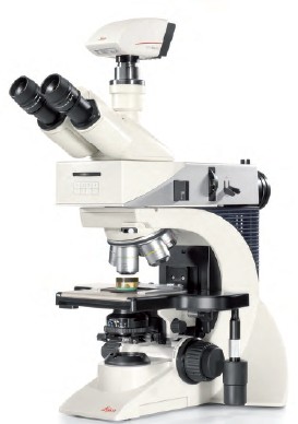
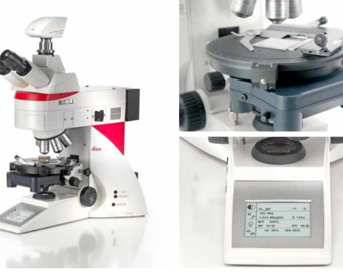
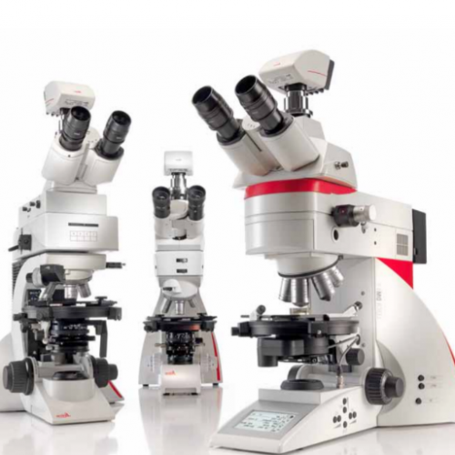 Depending on your application, you can choose from three different systems for polarization microscope. The Leica polarization microscope series is designed for all polarizing examinations: petrography, mineralogy, structure characterization, asbestos analysis, coal analysis (vitrinite reflection), and examination of liquid crystals.
 -
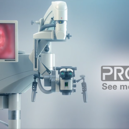
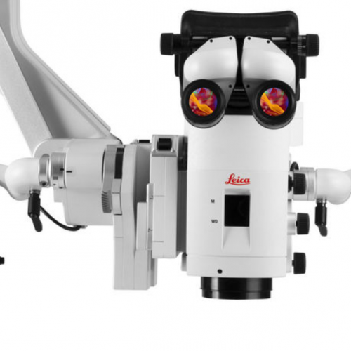
See more of the surgical site in one view so you can progress seamlessly through your next procedure with the PROvido floor stand surgical microscope. PROvido provides you with a bright, fully-focused view deeper into narrow cavities. A challenge that occurs daily in otolaryngology and spine procedures.
-
In delicate neurosurgery your vision and understanding of anatomical structures and physiological processes powers your decision-making. How would it impact outcomes if you had a single, precise, augmented view of the surgical field, in one augmented microscope platform?
-

-

-

-

-

-
-

-

-