-
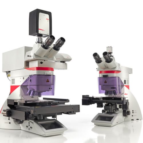 Laser Microdissection (LMD, also known as Laser Capture Microdissection or LCM) enables users to isolate specific single cells or entire areas of tissue. Powered by a unique laser design and dynamic software, Leica LMD systems allow users to easily isolate Regions of Interest (ROI) from entire areas of tissue down to single cells or even subcellular structures such as chromosomes.
 -
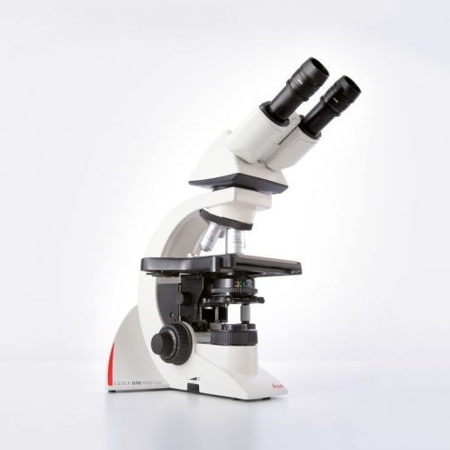 The Leica DM1000 LED features long-life LED illumination that provides near daylight, bright illumination with constant colour temperature and emits less heat.
 -
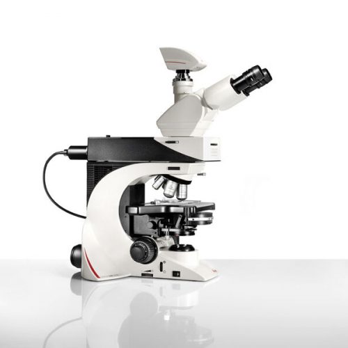
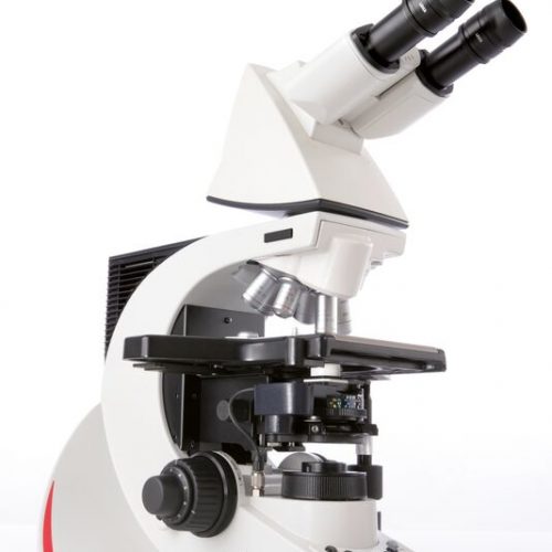
 The Leica DM2700 M flexible upright microscope system uses LED illumination for all contrast methods: brightfield (BF), darkfield (DF), differential interference contrast (DIC), qualitative polarization (POL), and fluorescence (FLUO) applications. It also offers built-in oblique illumination, which improves the visualization of surface topography and defects. Optionally, the Leica DM2700 M can also be equipped with a transmitted light axis. The Leica DM2700 M is equipped, e.g. with an N PLAN achromatic objective series with magnifications from 5x to 100x, a field of view of 22 mm, a flattened image field, and large working distances.
 -
 With their sophisticated modular design and high-performance fluorescence, the Leica DM2000 microscopes are ideal for complex tasks in pathology, cytology, and many other applications. For special diagnostics requirements, the microscope is certified for in-vitro-diagnostics (IVD) like in-vitro-fertilization (IVF).
 -
 Leica DM2500 & DM2500 LED optical microscopes are tools for demanding tasks in life science routine and research applications. With their transmitted light illumination, optical performance, and state-of-the-art accessories, they are especially well-suited for challenging life science research tasks that require differential interference contrast or high-performance fluorescence.
 -
 The Leica DM2700 M flexible upright microscope system uses LED illumination for all contrast methods: brightfield (BF), darkfield (DF), differential interference contrast (DIC), qualitative polarization (POL), and fluorescence (FLUO) applications. It also offers built-in oblique illumination, which improves the visualization of surface topography and defects. Optionally, the Leica DM2700 M can also be equipped with a transmitted light axis. The Leica DM2700 M is equipped, e.g. with an N PLAN achromatic objective series with magnifications from 5x to 100x, a field of view of 22 mm, a flattened image field, and large working distances.
 -
The Leica DM2700 M flexible upright microscope system uses LED illumination for all contrast methods: brightfield (BF), darkfield (DF), differential interference contrast (DIC), qualitative polarization (POL), and fluorescence (FLUO) applications. It also offers built-in oblique illumination, which improves the visualization of surface topography and defects. Optionally, the Leica DM2700 M can also be equipped with a transmitted light axis. The Leica DM2700 M is equipped, e.g. with an N PLAN achromatic objective series with magnifications from 5x to 100x, a field of view of 22 mm, a flattened image field, and large working distances.
-
 The Leica DM3000 microscopes for pathology, cytology, haematology and many other applications feature a motorized nosepiece, condenser head, automated light intensity adjustment, and optional foot pedal. These intuitive microscopes improve workflows significantly. For special diagnostics requirements, the microscope is certified for in-vitro-diagnostics (IVD) like in-vitro-fertilization (IVF).
 -
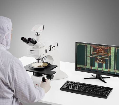 System for Microelectronics and Semiconductor. With a large field of view, the DM3 XL inspection system allows your team to identify defects faster and increase your yield rate. Make use of the 30% increased field of view of the unique macro objective. The DM3 XL uses LED illumination for all contrast methods. LED illumination provides a constant color temperature and offers real color imaging at all intensity levels.
 -
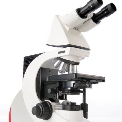
 Increase your work efficiency with the Leica DM4 B or Leica DM6 B upright digital research microscopes! Both are ideally suited to making your work life easier in biomedical research and clinical labs alike. You can configure the microscope that fits your need and budget with the Leica DM4 B and Leica DM6 B. Optimize your system to your application with different contrast methods and automated functions.
 -
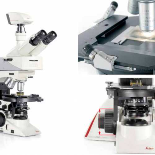
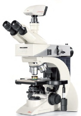
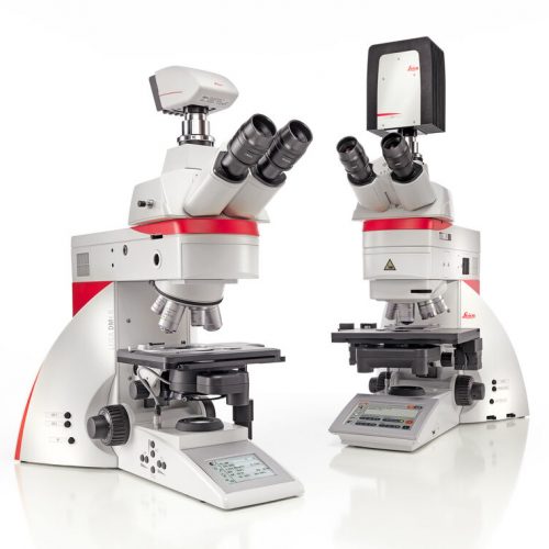
 Leica DM4 M and DM6 M digital microscopes for materials science and quality control offer truly reproducible microscopy, incredible optics and high-quality images. Store and recall your imaging conditions with a touch of a button. High quality microscope images make challenging inspection, measurement, and analysis tasks easy. Use the Leica DM4 M for manual routine inspection or the Leica DM6 M for fully automated materials analysis.