-
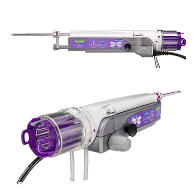 The Mammotome Revolve™ device is designed for accuracy, repeatability and efficiency, while staying true to your core purpose – acquiring the best quality tissue during your patient’s biopsy.
 -
Provides the speed and ease of a core needle with the advantages of vacuum-assisted technology.
Designed for procedural efficiency and an accurate sample.
-
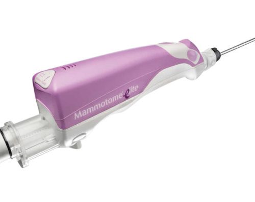
Offering clinicians the most comprehensive portfolio available, providing a customized solution for every breast biopsy need.
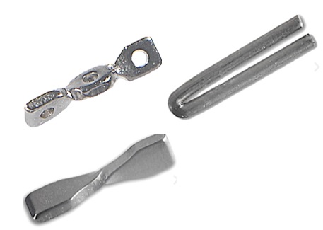
Breast marking technologies designed to meet your patients’ individual needs.
-
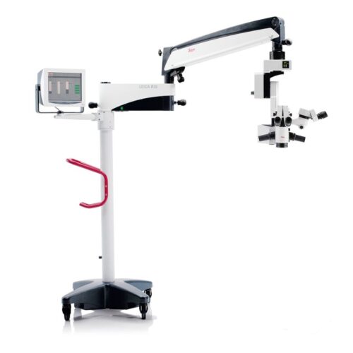
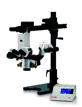
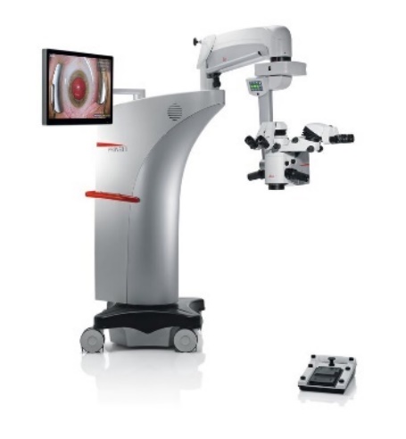 The Proveo 8 ophthalmic microscope provides the exact image you need at each moment of your procedure. Like a precision timepiece every element of the Proveo 8 microscope interconnects and works in perfect synchrony to optimize your view.
 -
 The Leica M844 F40 belongs to the premium class of ophthalmic surgical microscopes and offers optimal solutions particularly for posterior and high-end anterior segment surgery.
 -
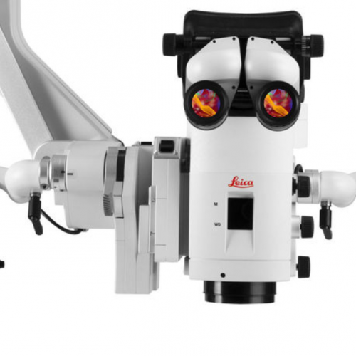
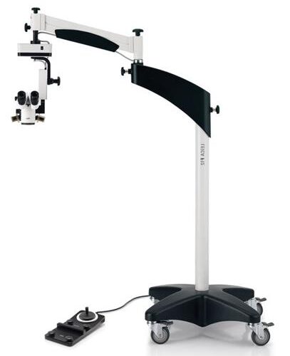
 The Leica M822 surgical microscope, with enhanced Red Reflex, meets the highest professional requirements of ophthalmic surgeons to perform cataract surgery more precisely and efficiently.
 -
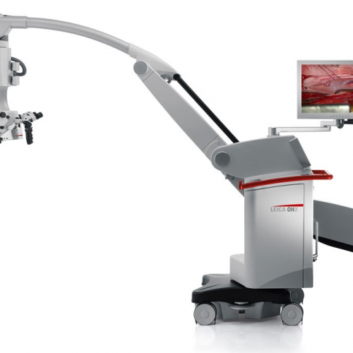
 The Leica OH4 surgical microscope provides superior reach, height, and clearance. The Leica OH4 floor stands that moves easily and when the brakes are released, the microscope stabilizes in only 1.5 seconds and remains vibration-free. By integrating new glass, glass coatings, and design parameters, high-resolution APO OptiChrome technology delivers an expanded working distance and greater depth of focus. In combination with the innovative light management systems BrightCare and AutoIris, you can operate and safe light levels and still see more than ever before. The M525 OH4 also offers FL800 and FL400 fluorescence for intraoperative visualization of blood flow and tumour tissue.
 -
Compact surgical microscope Smooth, fine movements, Xenon illumination systems, Motorized MultiFoc lens
The Leica M525 F20 surgical microscope combines brilliant optical quality with superior maneuverability. The crisp, sharp images and large depth of field allow the surgeon to see precise details. The working-distance-based illumination provides enough light for deep operative sites while supporting patient safety. Designed as an otolaryngology microscope, the Leica M525 F20 is also ideal for spine, dental, hand, and plastic/reconstructive procedures. Ease of movement -
Precision Surgical Microscope For Neurosurgery, Spine & Plastic Reconstructive Surgery
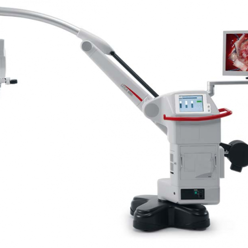
Precision comes standard in our M530 OHX, helping you achieve the highest standards of precision in your surgery. -
Neurosurgery Microscope
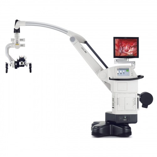
The Leica M530 OH6 takes image quality to a new level: FusionOptics, the groundbreaking technology by Leica Microsystems, unites an enhanced depth of field with high resolution to create an optimal view. FusionOptics also helps you to streamline your work: A larger area in full focus means that there is less need to refocus the microscope. FusionOptic let you stay focused – in every sense of the word. -
Premium Surgical Microscope for Microsurgery
The Leica M720 OH5 is the premium operating microscope for neurosurgery, otolaryngology and reconstructive microsurgery. With this operating microscope, surgeons will benefit from greater visibility in deep cavities or lesions and improved ergonomics and comfort. The Leica M720 OH5 surgical microscope also offers full HD 3D and 2D visualization and recording, so that the entire surgical team as well as students will be able to observe and learn more easily. The robust OH5 floor stand, designed by Mitaka, features long reach and electromagnetic brakes for lightweight positioning and fast stabilization. -
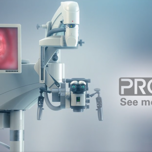
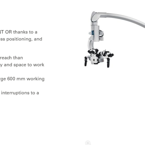
See more of the surgical site in one view so you can progress seamlessly through your next procedure with the PROvido floor stand surgical microscope. PROvido provides you with a bright, fully-focused view deeper into narrow cavities. A challenge that occurs daily in otolaryngology and spine procedures.
-
In delicate neurosurgery your vision and understanding of anatomical structures and physiological processes powers your decision-making. How would it impact outcomes if you had a single, precise, augmented view of the surgical field, in one augmented microscope platform?
-
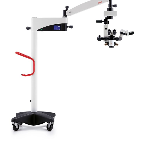 The Leica M620 F20 – an ophthalmic surgical microscope with its crisp, sharp OptiChrome™ optics gives the surgeon natural color, outstanding depth-of-focus and higher contrast for maximum detail recognition.
 -
 The DI C800 Digital Imaging Color Module is an easy-to-use and ergonomic solution that allows the surgeon to view imaging data in the highest quality. It takes XGA signals from a variety of external sources and displays them in the surgeon’s eyepiece
 -
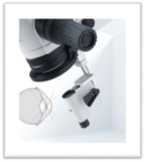
 The Leica Keratoscope is a ring illuminator used intraoperatively to qualitatively evaluate the corneal curvature of the eye for astigmatism. With the Leica Keratoscope the surgeon now has a cost-effective, integrated instrument to help assess the shape of the anterior surface of the cornea. This will aid him or her in making limbal relaxing incisions (LRIS) and positioning Toric IOLs.
 -
 The Leica RUV800 Retinal Upright Viewing system is designed to work with Leica ophthalmic microscopes. The Leica RUV800, with its integrated inverter, sits beneath the microscope’s optics and gives the surgeon, the assistant, and the video camera the same upright view of the retina. This makes it easier and safer to position and insert surgical instruments during surgery.
 -
 The Leica M220 F12 - an ophthalmic microscope is fully dedicated to the needs of ophthalmic surgery. Prestigious Leica optics motorized 5-step APO-chromatic magnification changer and focus, LED-illumination without fiber optics cables for direct and instant Red Reflex, and upgradeable XY-unit are standard features
 -
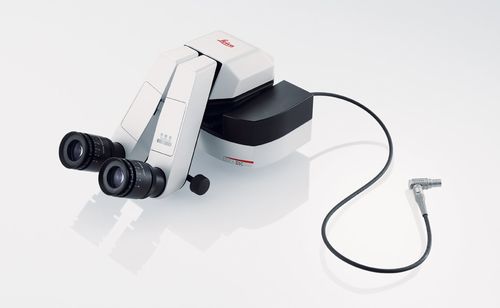
 High-performance Training Microscope The new Leica M620 TTS (tabletop stand) microscope meets the needs of surgical trainees as well as trainers. The Leica M620 TTS microscope system offers brilliant resolution, a large depth of field with outstanding stereopsis, and natural color reproduction on a convenient tabletop stand.
 -
You are always striving to reach the best possible patient outcome. To achieve this you need to see every detail even in narrow channels and to free maneuver yourself and your instruments.