-
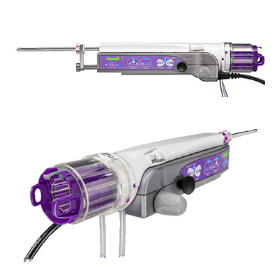 The Mammotome Revolve™ device is designed for accuracy, repeatability and efficiency, while staying true to your core purpose – acquiring the best quality tissue during your patient’s biopsy.
 -
Provides the speed and ease of a core needle with the advantages of vacuum-assisted technology.
Designed for procedural efficiency and an accurate sample.
-
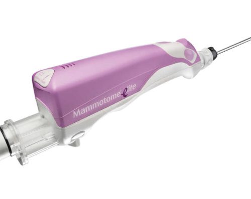
Offering clinicians the most comprehensive portfolio available, providing a customized solution for every breast biopsy need.
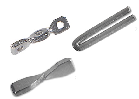
Breast marking technologies designed to meet your patients’ individual needs.
-
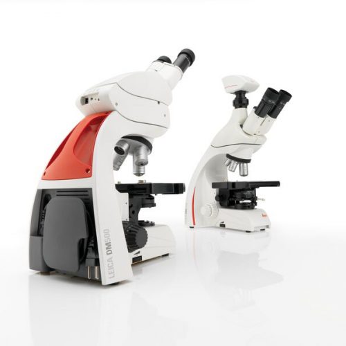 The Leica DM500 microscope with “plug and play” capability is the ideal tool to make teaching entry-level college and university Life Science courses easy and fun for the instructor and the student.
 -
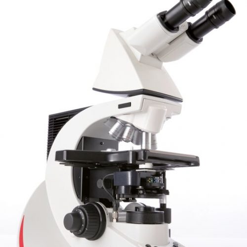
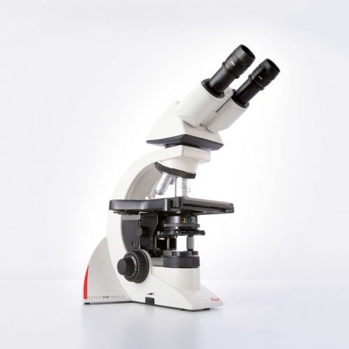 The Leica DM1000 LED features long-life LED illumination that provides near daylight, bright illumination with constant colour temperature and emits less heat.
 -
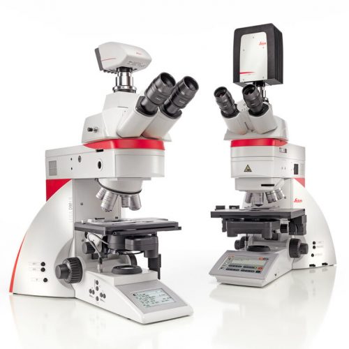
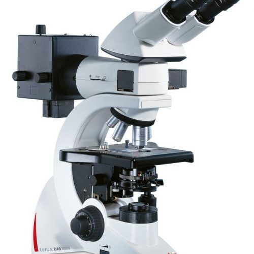
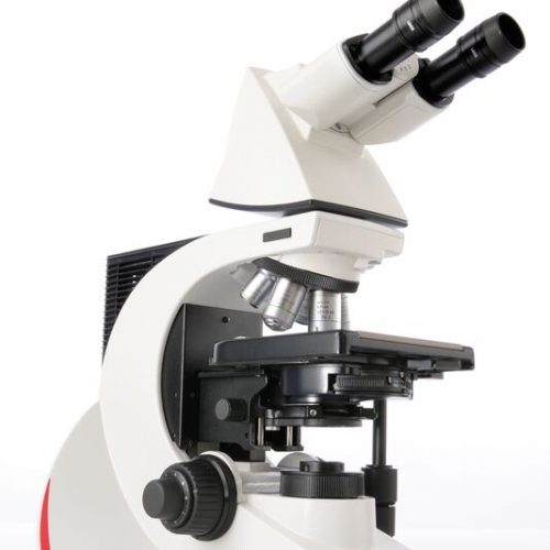 With their sophisticated modular design and high-performance fluorescence, the Leica DM2000 microscopes are ideal for complex tasks in pathology, cytology, and many other applications. For special diagnostics requirements, the microscope is certified for in-vitro-diagnostics (IVD) like in-vitro-fertilization (IVF).
 -
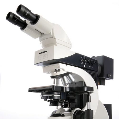
 Leica DM2500 & DM2500 LED optical microscopes are tools for demanding tasks in life science routine and research applications. With their transmitted light illumination, optical performance, and state-of-the-art accessories, they are especially well-suited for challenging life science research tasks that require differential interference contrast or high-performance fluorescence.
 -
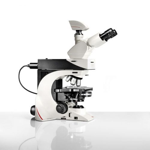
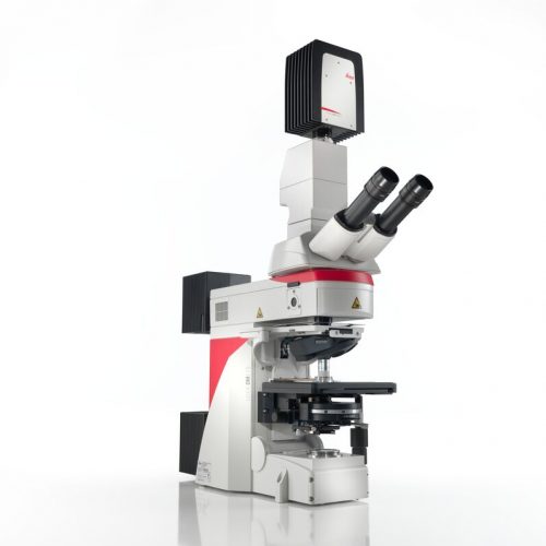
 The Leica DM3000 microscopes for pathology, cytology, haematology and many other applications feature a motorized nosepiece, condenser head, automated light intensity adjustment, and optional foot pedal. These intuitive microscopes improve workflows significantly. For special diagnostics requirements, the microscope is certified for in-vitro-diagnostics (IVD) like in-vitro-fertilization (IVF).
 -
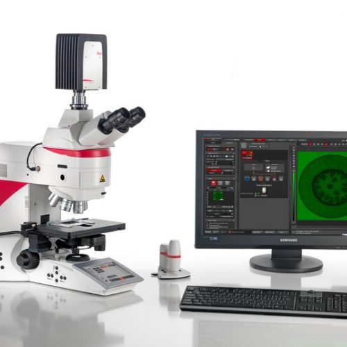
 Increase your work efficiency with the Leica DM4 B or Leica DM6 B upright digital research microscopes! Both are ideally suited to making your work life easier in biomedical research and clinical labs alike. You can configure the microscope that fits your need and budget with the Leica DM4 B and Leica DM6 B. Optimize your system to your application with different contrast methods and automated functions.
 -
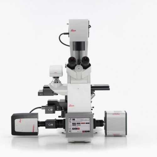
 Do you work in a field such as electrophysiology, evolutionary biology, or neuroscience where outstanding stability is required for successful experiments? The Leica DM6 FS fixed stage fluorescence microscope is a suitable instrument for your present and future challenges.
 -
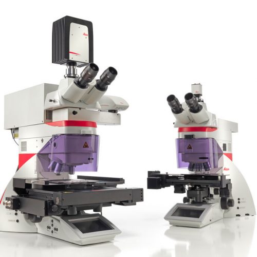 Laser Microdissection (LMD, also known as Laser Capture Microdissection or LCM) enables users to isolate specific single cells or entire areas of tissue. Powered by a unique laser design and dynamic software, Leica LMD systems allow users to easily isolate Regions of Interest (ROI) from entire areas of tissue down to single cells or even subcellular structures such as chromosomes.
 -
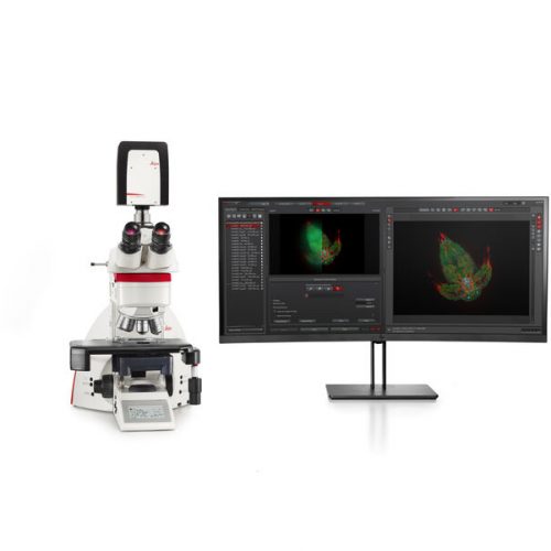 The THUNDER Imager Tissue allows real-time fluorescence imaging of 3D tissue sections typically used in neuroscience and histology research. Acquire rich, detailed images of thick tissues free of haze from out-of-focus blur.
 -
 Fluorescence is one of the most commonly used physical phenomena in biological and analytical microscopy, mainly because of its high sensitivity and high specificity. Fluorescence is a form of luminescence.
 -
 Fluorescence is one of the most commonly used physical phenomena in biological and analytical microscopy, mainly because of its high sensitivity and high specificity. Fluorescence is a form of luminescence.
 -
Fluorescence is one of the most commonly used physical phenomena in biological and analytical microscopy, mainly because of its high sensitivity and high specificity. Fluorescence is a form of luminescence.
-
 Fluorescence is one of the most commonly used physical phenomena in biological and analytical microscopy, mainly because of its high sensitivity and high specificity. Fluorescence is a form of luminescence.
 -
 Fluorescence is one of the most commonly used physical phenomena in biological and analytical microscopy, mainly because of its high sensitivity and high specificity. Fluorescence is a form of luminescence.
 -
 Fluorescence is one of the most commonly used physical phenomena in biological and analytical microscopy, mainly because of its high sensitivity and high specificity. Fluorescence is a form of luminescence.
 -
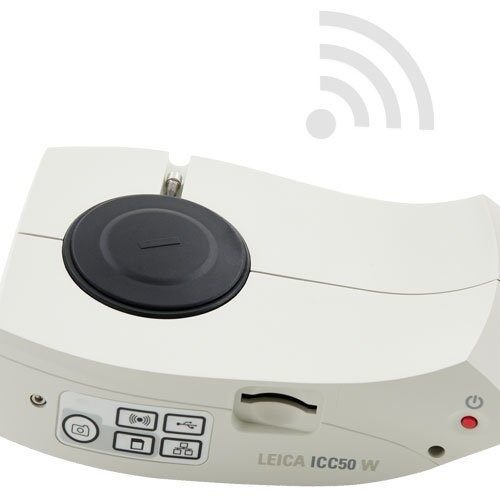 The ICC50 E exclusively uses the facilities network (WLAN or LAN) to allow students to connect to the camera. This is an ideal solution if you don`t want to add additional Wi-Fi access points to your existing wireless network concept. These 5-megapixel cameras can live-stream HD images to students’ smartphones or tablets. Students can connect to the ICC50 W either through their own internal Wi-Fi signal or through the facilities network.
 -
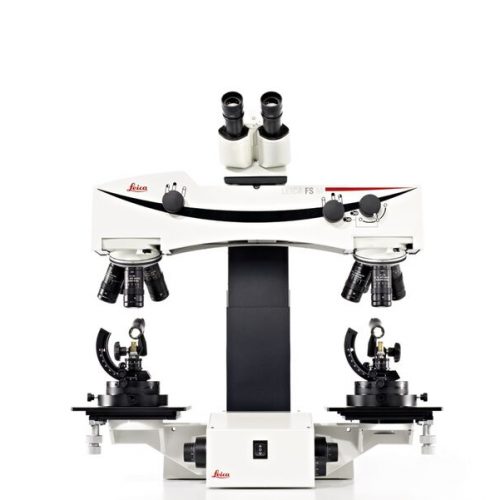 The Leica FS M manually-controlled forensic macroscope provides the flexibility, convenience, and comfort that make it the universal instrument of firearms and tool mark examiner. The versatile system is ideal for the simultaneous observation of evidence for training and consultation. The high-quality comparison bridge supports two precisely matched sets of apochromatically corrected objectives on quintuple ball-bearing nosepieces.
 -
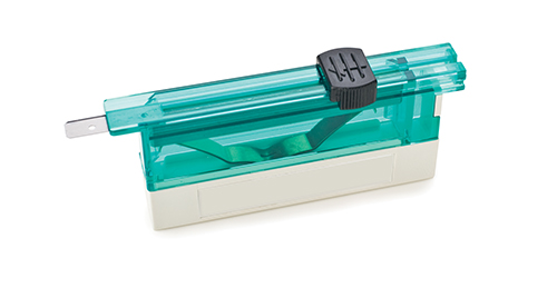
 When your application calls for thicker sections, look to the high-profile disposable blades DB80HS with sharpness, longevity and consistency.
 -
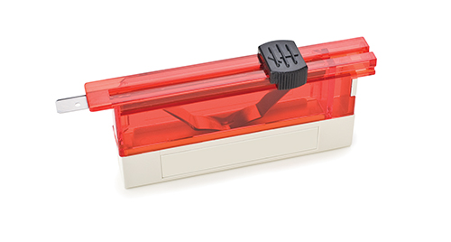
 The proprietary nature of the low-profile disposable blades DB80LX aids in cutting high quality, thin sections with even very difficult to section tissue including dense specimens. With select materials, sophisticated coating and an advanced profile, this blade quickly and efficiently allows you to section routine specimens, biopsies and dense tissues.
 -
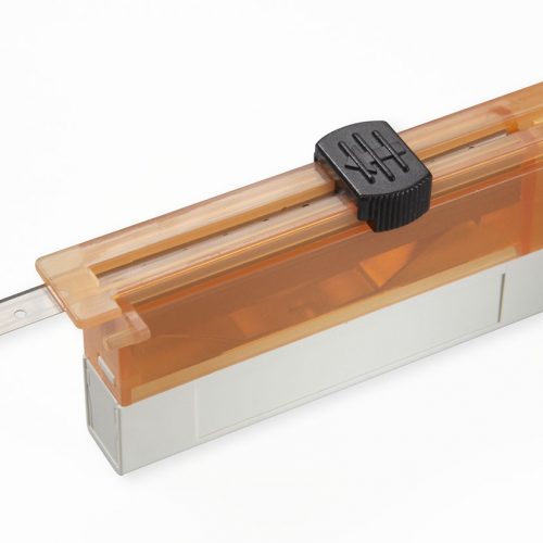
 Cut the very thin sections needed for diagnostic clarity. The low-profile disposable blades DB80LS provide a new level of quality with a low-profile shape, a premium finish and edge-to-edge, blade-to-blade consistency.
 -
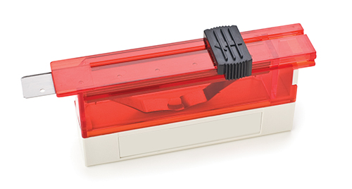
 Low-profile 819 disposable blades are 80 mm long x 8 mm high x 0.25 mm thick. Made of stainless steel, the blades are ultra-fine yet durable and specifically designed for use with Leica Biosystems microtomes or cryostats classified and labelled as in vitro diagnostic medical devices for histological medical diagnosis, e.g. cancer diagnosis.