-
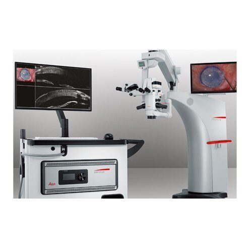 See even more: EnFocus with Proveo 8 Combine EnFocus OCT with the premium Proveo 8 microscope for a complete visualization platform for all your ophthalmic surgeries.
 -
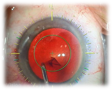 You strive to deliver minimal residual astigmatism so your patient can be glasses-free after cataract surgery. When dealing with miniscule eye structures, there is no room for inaccuracy.
 -
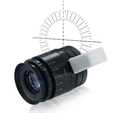 The Leica ToricEyePiece is an ocular with a rotatable angle scale helping the surgeon to position and align toric intraocular lenses (IOL) efficiently, ensuring optimal correction of astigmatism. EASY TO USE Simply rotate the graticule using the sterile handle and identify the desired target position for the toric IOL
 -
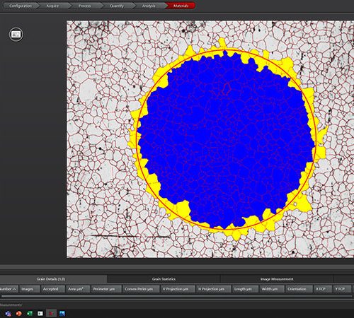 For industrial quality control and materials research, your goal is to deliver accurate and reliable results. You need software that enables you to work with speed, efficiency, and precision and facilitates your daily tasks.
 -
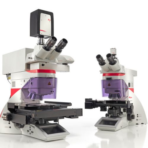 Laser Microdissection (LMD, also known as Laser Capture Microdissection or LCM) enables users to isolate specific single cells or entire areas of tissue. Powered by a unique laser design and dynamic software, Leica LMD systems allow users to easily isolate Regions of Interest (ROI) from entire areas of tissue down to single cells or even subcellular structures such as chromosomes.
 -
 The intuitive A60 F and A60 S stereo microscopes fulfill what you need – high sample throughput, optimum visibility of product details and components, and easy processing of subassemblies.
 -
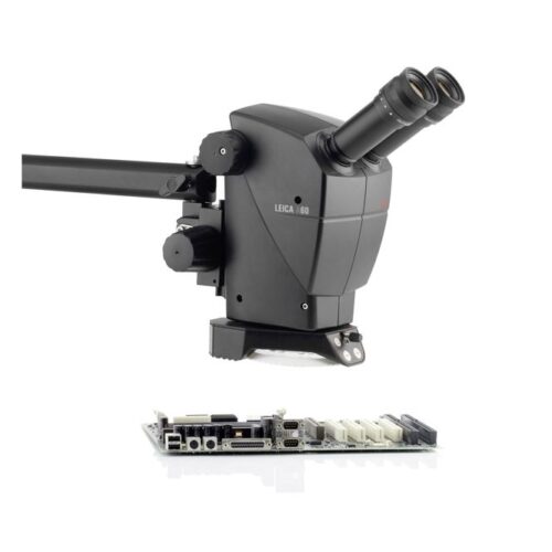 The intuitive A60 F and A60 S stereo microscopes fulfill what you need – high sample throughput, optimum visibility of product details and components, and easy processing of subassemblies. The Leica A60 stereomicroscope does not require the user to be a microscope expert. The components of the preconfigured system are perfectly matched to each other to provide easy operation with the best optical results. Choose between two different stands: the Leica A60 F with flex arm for maximum freedom of movement or the Leica A60 S with swing arm for high stability.
 -
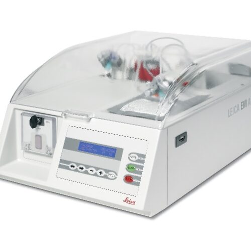 The Leica EM AC20 automatic contrasting system for ultrathin sections ensures minimum user contact with reagents and reduced reagent consumption.
 -
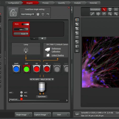 The Leica Application Suite (LAS) integrates Leica automated microscopes and digital cameras and provides one common, easy-to-use, consistent user interface.
 -
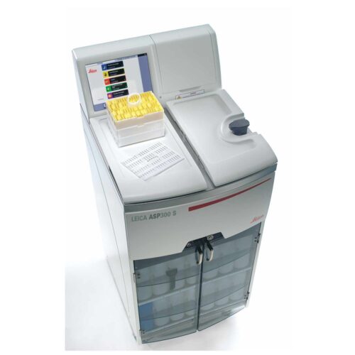 Designed for routine and research histopathology of up to 300 cassettes. Proven technology combined with top quality components. Optional RemoteCare diagnostic provide superior instrument reliability. Simplify user operations with an intuitive user interface, color touch screen and a variety of "smart" features. Improve specimen quality and laboratory economy with the Reagent Management System and quick start for commonly used programs.
 -
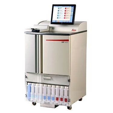 Focus on what’s critical: Quality, specimen safety, productivity. The ASP6025 supports diagnostic clarity thanks to an optimized reagent management system and validated processing protocols. Built-in safety features and reagent quality control deliver specimen protection and accurate processing results.
 -
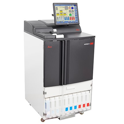 The New ASP6025 S is designed to improve your lab’s performance with quality-driven technology that optimizes tissue processing performance and supports rapid processing. Designed for increased performance and reliability, the new ASP6025 S processes up to 300 cassettes simultaneously for faster turnaround times.
 -
 The Leica CM1520 is the best-value cryostat available for cryosectioning including critical applications such as Mohs surgery. The Leica CM1520 delivers high-quality sections the fast, simple and efficient way. Time is crucial when sectioning frozen specimens. An actively cooled quick freezing shelf with defrost function and a powerful refrigeration system are provided to get rapid results.
 -
 Leica CM1950 is the cryostat for Standard Applications in the Clinical Histopathology Laboratory. It is your perfect assistant for high-quality, fast and safe sectioning. This high-throughput cryostat delivers diagnostic confidence by reliably producing quality sections to help provide an accurate diagnosis - even with the most complicated tissue types.
 -
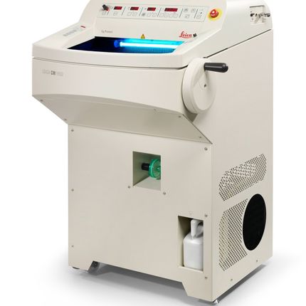
 The Leica CM1860 UV, developed with an emphasis on diagnostic confidence, safety and ergonomics, is the cryostat for histopathology laboratory applications. The high-throughput cryostat creates diagnostic confidence by reliably producing quality sections that help provide an accurate diagnosis.
 -
 Leica CM1950 cryostat design is the result of extensive customer consultations.
 -
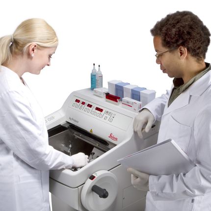
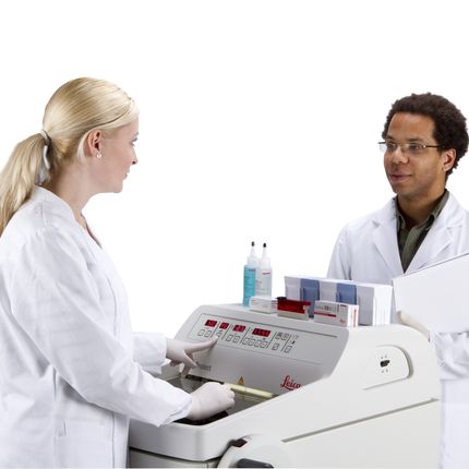
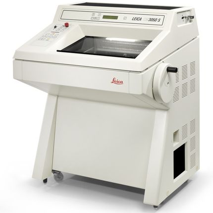 Primarily designed for the demanding needs of cryosectioning in biomedical, neuro-anatomical and pharmaceutical research. The Leica CM3050 S cryostat features superior user comfort with excellent safety standards for practically all types of cryosectioning applications. It is the instrument of choice for all research applications and for advanced clinical cryosectioning needs. Particularly when working with delicate specimens – for example brain samples in neuroscience – the precise specimen orientation and the specimen feed system guarantees reproducible, thin, serial sections of maximum quality.
 -
 The Leica CV5030 fully automated glass coverslipper produces slides with superior optical quality for reliable long-term storage. Its capability of handling a large variety of slide racks from different suppliers makes the Leica CV5030 very flexible. Most common mounting media, including xylene-free varieties, can be used. The operator can choose wet or dry coverslipping. The CV5030’s performance achieves three goals: Highly consistent and reliable coverslipping quality, full adaptation to individual laboratory set-ups, integration into staining/coverslipping workstations to form a fully automated, user-friendly operation system.
 -
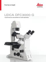 The Leica DFC3000 G is a grayscale USB 3.0 microscope camera for routine fluorescence applications. You will receive crisp images due to its unique passive cooling architecture and its highly sensitive CCD sensor. Correlated pixel double sampling together with a reduction of the ambient temperature of the sensor offers an exceptionally clear and noise -free signal. Its CCD sensor is particularly suitable for low light situations such as fluorescence and will capture even smallest amounts of light.
 -
 These cooled 2.8 megapixel cameras provide a new paradigm for camera image quality through truly innovative design. The monochrome DFC7000 GT for demanding fluorescence applications, or the color DFC7000 T developed for both brightfield and fluorescence imaging.
 -
 The Leica DFC9000 sCMOS camera will turn your imagination into reality. This monochrome microscope camera with newest scientific CMOS (sCMOS) sensor technology for fluorescence imaging enables you to image your cells under near-native conditions.
 -
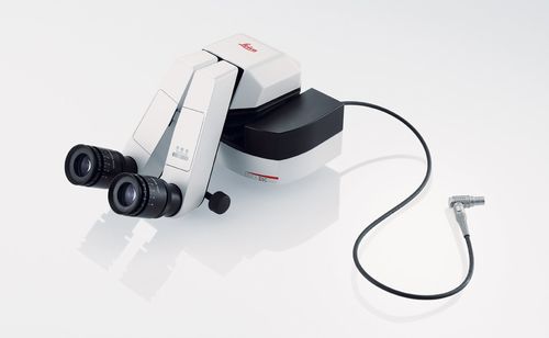 The DI C800 Digital Imaging Color Module is an easy-to-use and ergonomic solution that allows the surgeon to view imaging data in the highest quality. It takes XGA signals from a variety of external sources and displays them in the surgeon’s eyepiece
 -
 Fluorescence is one of the most commonly used physical phenomena in biological and analytical microscopy, mainly because of its high sensitivity and high specificity. Fluorescence is a form of luminescence.
 -
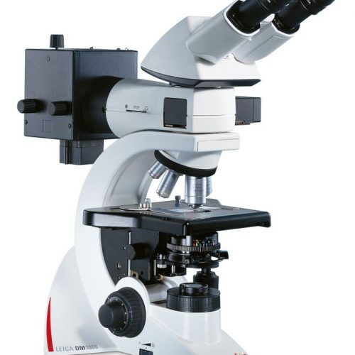
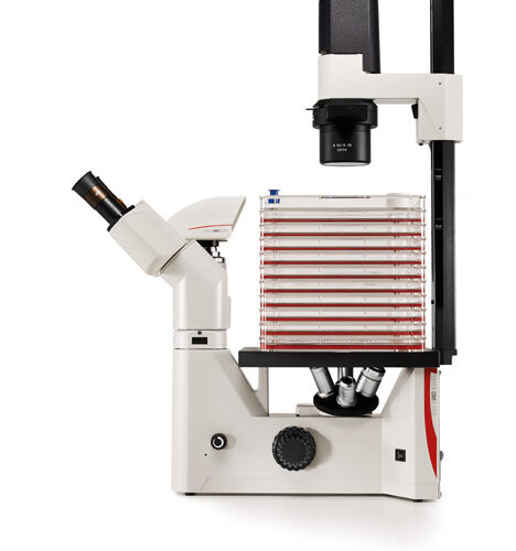 The Leica DM IL LED features a comprehensive set of contrast methods to monitor your specimen the way you need. High-quality Phase contrast, excellent modulation contrast and brilliant fluorescence are just one fingertip away. Robust stability, plenty of space to work with tools, long working distances to accommodate large culture flasks and a stable illumination without heat make work at the microscope easy and convenient.