-
 The Leica DM2700 M flexible upright microscope system uses LED illumination for all contrast methods: brightfield (BF), darkfield (DF), differential interference contrast (DIC), qualitative polarization (POL), and fluorescence (FLUO) applications. It also offers built-in oblique illumination, which improves the visualization of surface topography and defects. Optionally, the Leica DM2700 M can also be equipped with a transmitted light axis. The Leica DM2700 M is equipped, e.g. with an N PLAN achromatic objective series with magnifications from 5x to 100x, a field of view of 22 mm, a flattened image field, and large working distances.
 -
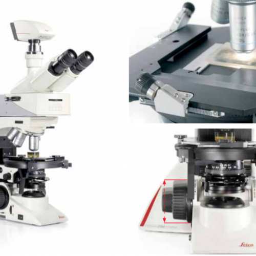
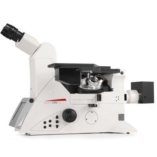
 The Leica DM2700 P offers a level of reproducibility that is one-of-a-kind in its class, the built-in focus stop protects your samples and the front lens of the objective. For samples of equal height, the focus stop makes the focusing plane easier to reconstruct so you can concentrate entirely on your application. Color-coded lenses match the color-coded field and aperture diaphragm adjustment (CDA), to ensure that the illumination conditions are always matched to the objective. Constant color temperature by advanced LED technology.
 -
 The Leica DM2700 M flexible upright microscope system uses LED illumination for all contrast methods: brightfield (BF), darkfield (DF), differential interference contrast (DIC), qualitative polarization (POL), and fluorescence (FLUO) applications. It also offers built-in oblique illumination, which improves the visualization of surface topography and defects. Optionally, the Leica DM2700 M can also be equipped with a transmitted light axis. The Leica DM2700 M is equipped, e.g. with an N PLAN achromatic objective series with magnifications from 5x to 100x, a field of view of 22 mm, a flattened image field, and large working distances.
 -
 The Leica DM3000 microscopes for pathology, cytology, haematology and many other applications feature a motorized nosepiece, condenser head, automated light intensity adjustment, and optional foot pedal. These intuitive microscopes improve workflows significantly. For special diagnostics requirements, the microscope is certified for in-vitro-diagnostics (IVD) like in-vitro-fertilization (IVF).
 -
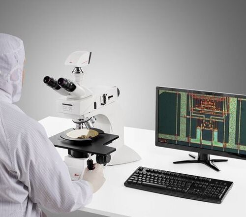 System for Microelectronics and Semiconductor. With a large field of view, the DM3 XL inspection system allows your team to identify defects faster and increase your yield rate. Make use of the 30% increased field of view of the unique macro objective. The DM3 XL uses LED illumination for all contrast methods. LED illumination provides a constant color temperature and offers real color imaging at all intensity levels.
 -
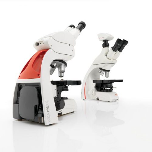
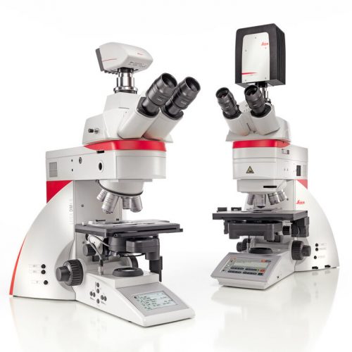 Increase your work efficiency with the Leica DM4 B or Leica DM6 B upright digital research microscopes! Both are ideally suited to making your work life easier in biomedical research and clinical labs alike. You can configure the microscope that fits your need and budget with the Leica DM4 B and Leica DM6 B. Optimize your system to your application with different contrast methods and automated functions.
 -
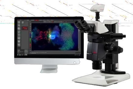
 Fluorescence is one of the most commonly used physical phenomena in biological and analytical microscopy, mainly because of its high sensitivity and high specificity. Fluorescence is a form of luminescence.
 -
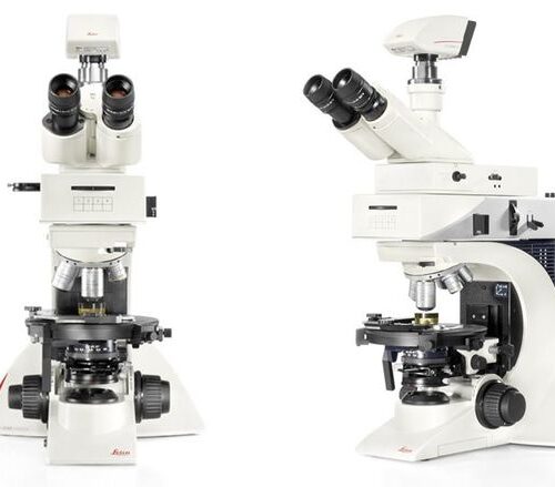
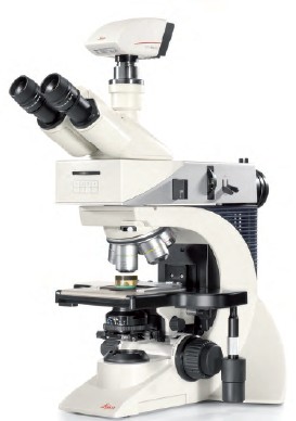
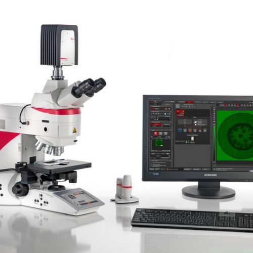
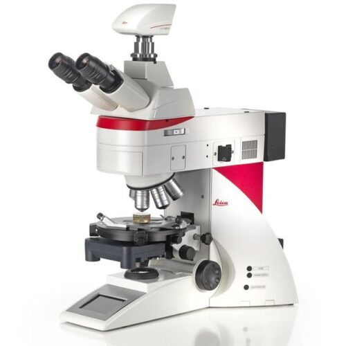
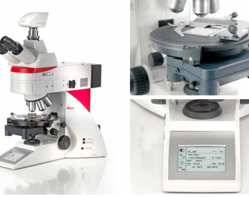
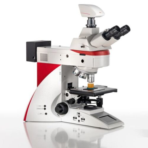
 Leica DM4 M and DM6 M digital microscopes for materials science and quality control offer truly reproducible microscopy, incredible optics and high-quality images. Store and recall your imaging conditions with a touch of a button. High quality microscope images make challenging inspection, measurement, and analysis tasks easy. Use the Leica DM4 M for manual routine inspection or the Leica DM6 M for fully automated materials analysis.
 -
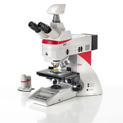
 Fully coded and semi-automated. The Leica DM4 P automatically detects which contrast method and objective are being used. This provides valuable consistency and reproducibility for your research. Manual diaphragm setting is no longer required, either in the transmitted light or incident light method. Light intensity automatically adjusts to the objective. Image brightness remains constant when switching objectives, which eliminates glare.
 -
 The Leica DM2700 M flexible upright microscope system uses LED illumination for all contrast methods: brightfield (BF), darkfield (DF), differential interference contrast (DIC), qualitative polarization (POL), and fluorescence (FLUO) applications. It also offers built-in oblique illumination, which improves the visualization of surface topography and defects. Optionally, the Leica DM2700 M can also be equipped with a transmitted light axis. The Leica DM2700 M is equipped, e.g. with an N PLAN achromatic objective series with magnifications from 5x to 100x, a field of view of 22 mm, a flattened image field, and large working distances.
 -
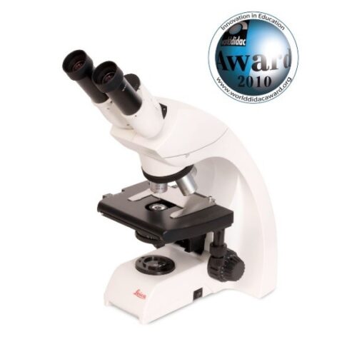
 The Leica DM500 microscope with “plug and play” capability is the ideal tool to make teaching entry-level college and university Life Science courses easy and fun for the instructor and the student.
 -
 LEICA DM500 Binocular Microscope, with 30 degree Bino EZ tube 10x/B, Plan Objectives (4x/0.1NA, 10x/0.22NA, 40x/0.65NA, Plan 100x/1.25NA) Inmersion oil, us power cord.
 -
 Do you work in a field such as electrophysiology, evolutionary biology, or neuroscience where outstanding stability is required for successful experiments? The Leica DM6 FS fixed stage fluorescence microscope is a suitable instrument for your present and future challenges.
 -
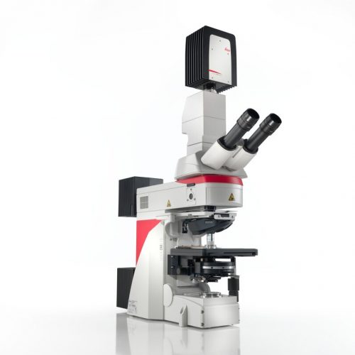
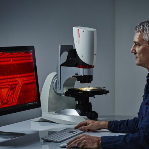
 The integrated laser spectroscopy function of the DM6 M LIBS delivers the chemical composition of the microstructure that you see in the microscope image - within a second. Identify the microstructure composition of interest, then trigger the LIBS analysis with a single click. The DM6 M LIBS 2-in-1 solution allows you to perform both structural and elemental/chemical analysis of material phases, e.g., minerals, alloys, ceramics, etc. There is no need for sample preparation nor transfer between 2 or more devices. The entire analysis workflow occurs with a single instrument.
 -
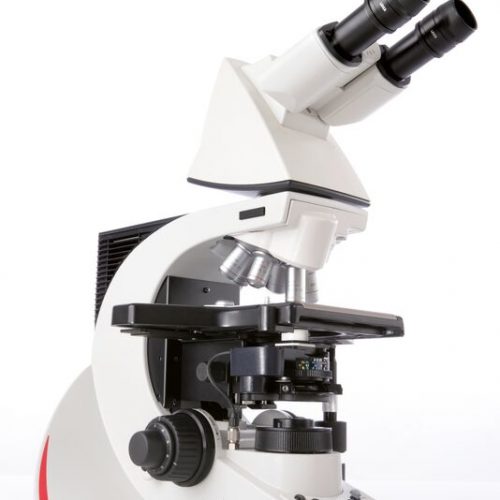
 The Leica DM750 M is the ideal microscope for basic materials applications in an Industrial Lab or Material Science course. It can be equipped with various specimen holders to accommodate mounted specimens of different diameters. The unique Reflected light LED illuminator provides brightfield, polarized light, and oblique illumination. This allows you to work with many different specimens with the same microscope configuration.
 -
 The Leica DM750 P is the ideal polarizing microscope for university and other instructional use, offering a standard and an advanced Bertrand lens module for unsurpassed ease of operation. The Leica DM750 P is the ideal polarizing microscope for university and other instructional use, offering a standard and an advanced Bertrand lens module for unsurpassed ease of operation.
 -
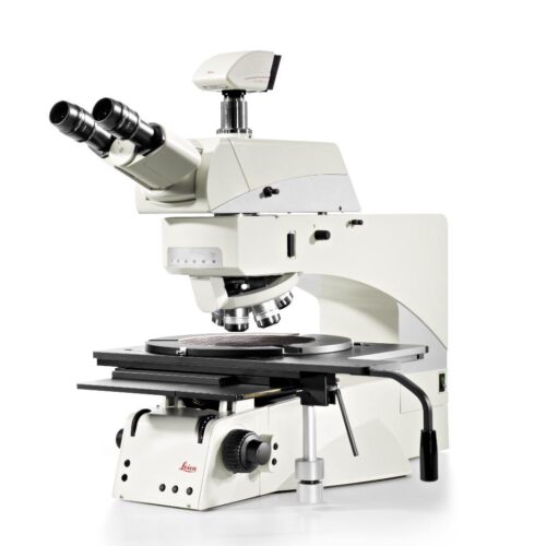 The Leica DM8000 M and Leica DM12000 M optical inspection systems provide an innovative yet cost-effective system solution for mastering present and future inspection challenges with confidence. Inspection, process control and defect analysis of wafers or LCDs and TFTs has to be fast, To detect macro defects, the Leica DM8000 M and DM12000 M have a micro/macro mode for rapid scanning of large components. The macro magnification captures an object field of approximately 40 mm.
 -
 The Leica DMC2900 is a digital USB 3.0 microscope camera with a 3.1 Megapixel CMOS sensor. It is an ideal tool for brightfield microscopic standard applications in research, life science and industry that often require capturing, documenting, and analyzing color images at optimum visibility of microstructures in little time. The Leica DMC2900 is a digital USB 3.0 microscope
 -
 Leica DMC4500 color camera. It has been designed as a versatile, uncomplicated tool that simplifies the imaging process from capture through to processing. It is ideally suited to life science applications such as documenting slides and organisms, pathology or pharmaceutical testing and industrial applications such as quality control and failure analysis.
 -
 The DMC5400 color CMOS camera offers high frame rate & high-resolution color images even at low magnification. It is optimized to produce fast, high quality images for documentation, evaluation and analysis, for a wide variety of samples and applications in manufacturing and life science research.
 -
 The DMC6200 camera delivers amazing images with intense color detail and contrast - whether at the lowest or highest magnification. Its state-of-the-art CMOS sensor has a 5.86 μm pixel size, 2.3 megapixels sensor resolution, and an astounding dynamic range of 73 dB (4000:1). Achieve an image resolution of up to 20.7 megapixels with cutting-edge pixel shift technology.
 -
 Designed to help you maximize efficiency, Leica DCM8 unites the advantages of High Definition confocal microscopy with interferometry into one versatile, dual-core system. Ultra-fast analysis is ensured thanks to one-click mode selection, sophisticated software and HD confocal scanning without moving parts.
 -
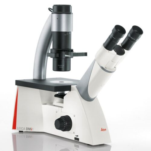 the Leica DMi1 the perfect partner for your live cell laboratory. Use the S40 condenser for optimal resolution with slides, petri dishes, multi-well dishes, and most types of flasks: The S40 provides an extra 10 mm of free space by simply moving it up. The Leica DMi1 camera version is an all-in-one solution for image capturing and cell culture documentation. You can choose between 2.5- or 5.0-megapixels resolution. The camera is mounted at the back of the stand. No additional trinocular tube is needed.
 -
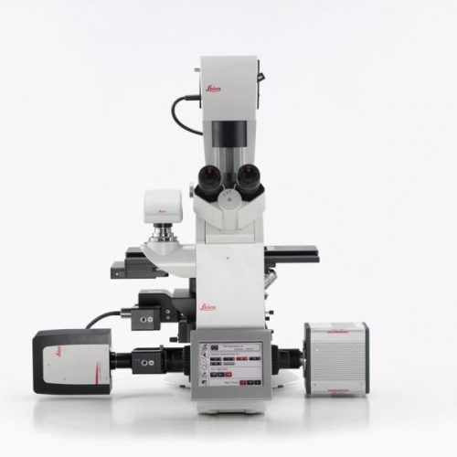
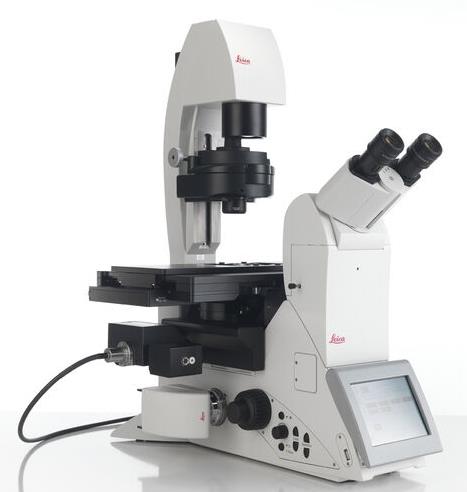 The modular DMi8 inverted microscope is the heart of the DMi8 S platform solution. For routine to live cell research, the DMi8 S platform is a complete solution. Whether you need to precisely follow the development of a single cell in a dish, screen through multiple assays, obtain single molecule resolution, or tease out behaviors of complex processes, a DMi8 S system will enable you to see more, see faster, and find the hidden.
 -
 Fluorescence is one of the most commonly used physical phenomena in biological and analytical microscopy, mainly because of its high sensitivity and high specificity. Fluorescence is a form of luminescence.
 -
 The modular DMi8 inverted microscope is the heart of the DMi8 S platform solution. For routine to live cell research, the DMi8 S platform is a complete solution. Whether you need to precisely follow the development of a single cell in a dish, screen through multiple assays, obtain single molecule resolution, or tease out behaviors of complex processes, a DMi8 S system will enable you to see more, see faster, and find the hidden.
 -
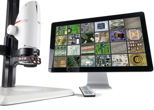
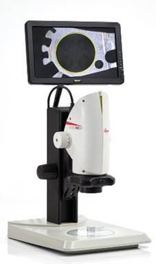 Digital microscope system for digital inspection, observation and measurement. From tiniest detail to an overview, the optics ensures magnification of up to 300x. The built-in HDMI microscope camera provides full high definition live images of up to 30fps and a resolution of 5Mpixels.
 -
 Digital microscope system for digital inspection, observation and measurement. From tiniest detail to an overview, the optics ensures magnification of up to 300x. The built-in HDMI microscope camera provides full high definition live images of up to 30fps and a resolution of 5Mpixels.
 -
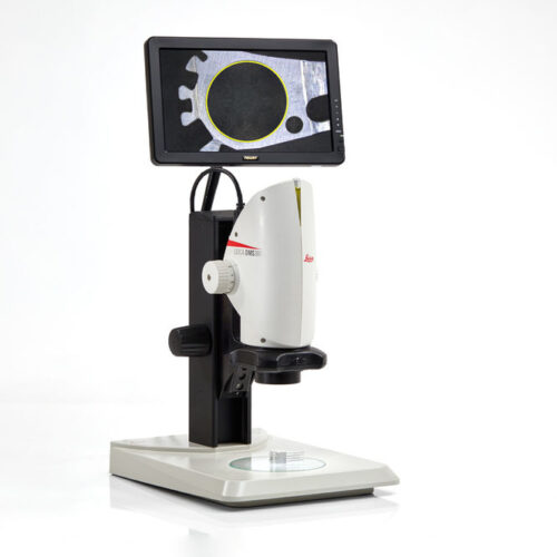 The Leica DMS300 is a complete digital microscope system that utilizes a HDMI-monitor instead of eyepieces. Leica’s high quality 8:1 zoom optics are combined with a 2.5MP camera to provide a full high-definition live image with up to 30fps. The Leica DMS300 produces high-quality, full-color still images as well as Full-HD movies as a standalone system.
 -
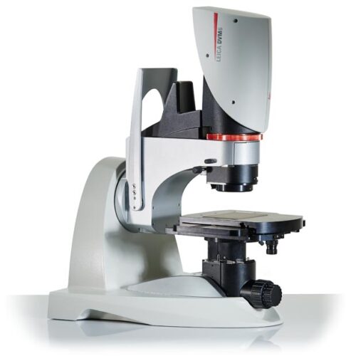
 With the DVM6, you can get from the big picture to smallest details in an instant. You can seamlessly carry on working even if changing objective is required, as the sample always stays in focus and no pre-adjustments are needed. With the tilting function you can observe your sample from different angles from up to ±60° With the DVM6, you can get from the big picture to smallest details in an instant. You can seamlessly carry on working even if changing objective is required, as the sample always stays in focus and no pre-adjustments are needed. With the tilting function you can observe your sample from different angles from up to ±60°.
 -
 With the DVM6, you can get from the big picture to smallest details in an instant. You can seamlessly carry on working even if changing objective is required, as the sample always stays in focus and no pre-adjustments are needed.
 -
 If you work in quality control/assurance, failure analysis, research and development, or in forensics, searching for the detail can take up a lot of your time in microscopy. The DVM6 digital microscope is a fast, reliable and easy to use solution that combines outstanding optics, intuitive operation, and smart software to save you time.
 -
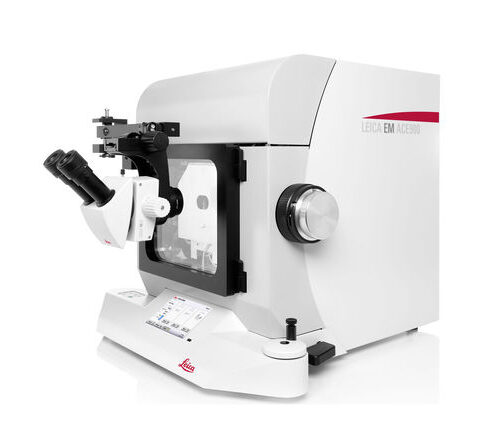 The Leica EM ACE900 is a high-end sample preparation system for freeze fracturing, freeze etching and high resolution cryo coating. This instrument is easy and intuitive to use.
 -
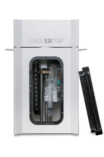 Freeze Substitution and Low Temperature Embedding System The Leica EM AFS2 performs freeze substitution and progressive lowering of temperature (PLT) techniques and allows low temperature embedding and polymerization of resins.
 -
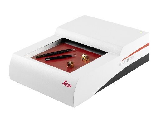 During Cryo preparation, tools such as forceps and sample holders often ice over and cannot be immediately used for the next sample run. De-icing and drying of the tools at room temperature is time consuming and can cause damage to sensitive components and may prevent their operation.