-
 The Leica DM1750 M is a material microscope designed for rapid, accurate analysis results even for a use in rough ambient conditions. Working with the Leica DM1750 M you will see; how simple and reliable microscopy can be. Its robust design contains an excellent optical system and allows the inspection even of larger samples, in brightfield, oblique- or with polarized light. The entire reflected light illumination is carried out with Power-LEDs which allow an inspection with different illumination angles, especially suitable for the detection of micro scratches or for gaining height information.
 -
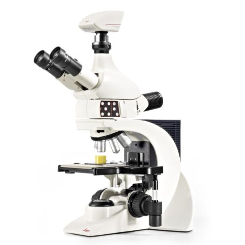
 The Leica DM2700 M flexible upright microscope system uses LED illumination for all contrast methods: brightfield (BF), darkfield (DF), differential interference contrast (DIC), qualitative polarization (POL), and fluorescence (FLUO) applications. It also offers built-in oblique illumination, which improves the visualization of surface topography and defects. Optionally, the Leica DM2700 M can also be equipped with a transmitted light axis. The Leica DM2700 M is equipped, e.g. with an N PLAN achromatic objective series with magnifications from 5x to 100x, a field of view of 22 mm, a flattened image field, and large working distances.
 -
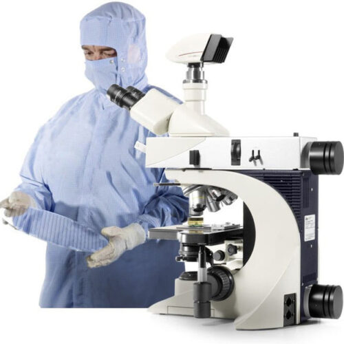
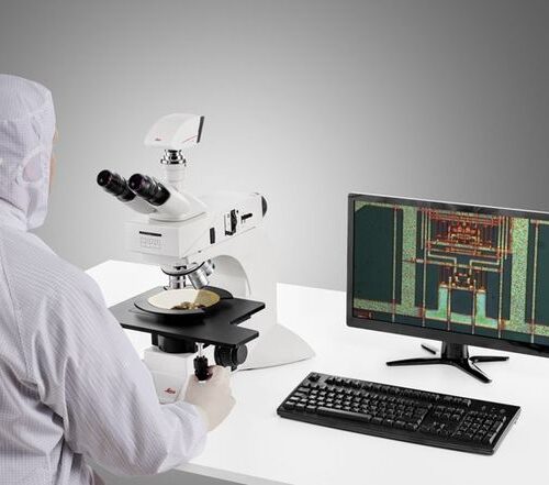
 Leica DM4 M and DM6 M digital microscopes for materials science and quality control offer truly reproducible microscopy, incredible optics and high-quality images. Store and recall your imaging conditions with a touch of a button. High quality microscope images make challenging inspection, measurement, and analysis tasks easy. Use the Leica DM4 M for manual routine inspection or the Leica DM6 M for fully automated materials analysis.
 -
 The Leica DM750 M is the ideal microscope for basic materials applications in an Industrial Lab or Material Science course. It can be equipped with various specimen holders to accommodate mounted specimens of different diameters. The unique Reflected light LED illuminator provides brightfield, polarized light, and oblique illumination. This allows you to work with many different specimens with the same microscope configuration.
 -
 System for Microelectronics and Semiconductor. With a large field of view, the DM3 XL inspection system allows your team to identify defects faster and increase your yield rate. Make use of the 30% increased field of view of the unique macro objective. The DM3 XL uses LED illumination for all contrast methods. LED illumination provides a constant color temperature and offers real color imaging at all intensity levels.
 -
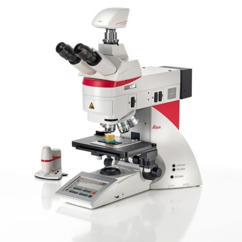
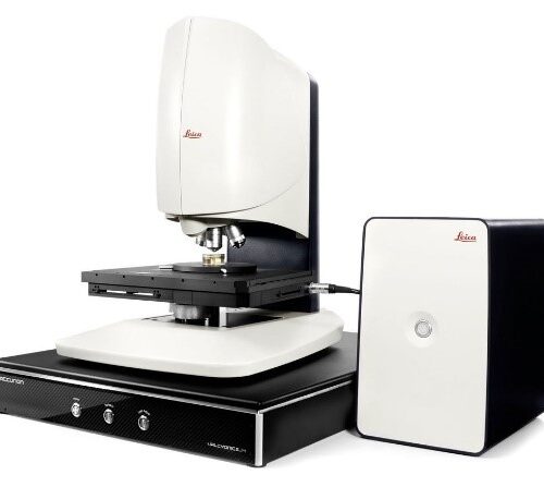 Designed to help you maximize efficiency, Leica DCM8 unites the advantages of High Definition confocal microscopy with interferometry into one versatile, dual-core system. Ultra-fast analysis is ensured thanks to one-click mode selection, sophisticated software and HD confocal scanning without moving parts.
 -
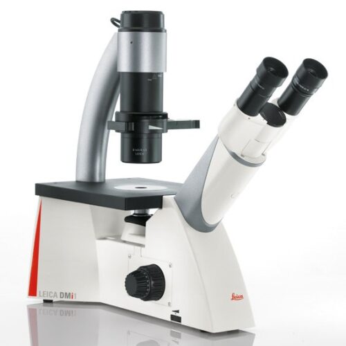 the Leica DMi1 the perfect partner for your live cell laboratory. Use the S40 condenser for optimal resolution with slides, petri dishes, multi-well dishes, and most types of flasks: The S40 provides an extra 10 mm of free space by simply moving it up. The Leica DMi1 camera version is an all-in-one solution for image capturing and cell culture documentation. You can choose between 2.5- or 5.0-megapixels resolution. The camera is mounted at the back of the stand. No additional trinocular tube is needed.
 -
 The Leica DM IL LED features a comprehensive set of contrast methods to monitor your specimen the way you need. High-quality Phase contrast, excellent modulation contrast and brilliant fluorescence are just one fingertip away. Robust stability, plenty of space to work with tools, long working distances to accommodate large culture flasks and a stable illumination without heat make work at the microscope easy and convenient.
 -
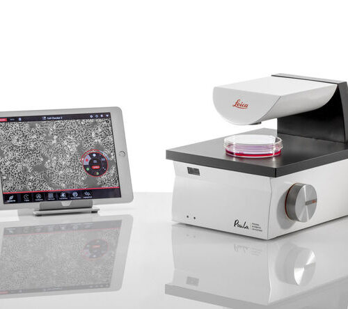 PAULA helps you to speed up your daily cell culture tasks and standardize the results to improve your downstream workflow, offering you essential analysis apps like confluence and transfection efficiency check.
 -
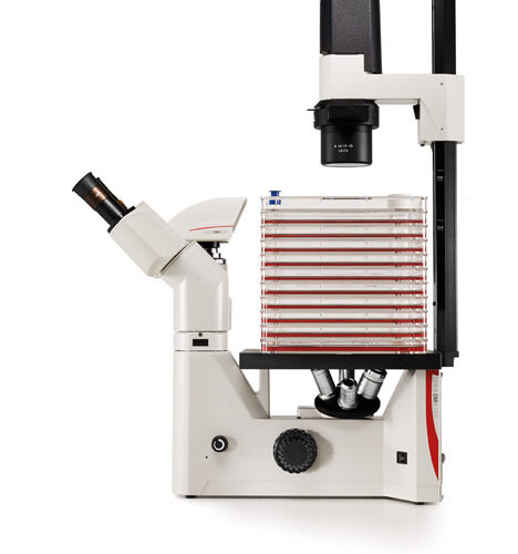
 The modular DMi8 inverted microscope is the heart of the DMi8 S platform solution. For routine to live cell research, the DMi8 S platform is a complete solution. Whether you need to precisely follow the development of a single cell in a dish, screen through multiple assays, obtain single molecule resolution, or tease out behaviors of complex processes, a DMi8 S system will enable you to see more, see faster, and find the hidden.
 -
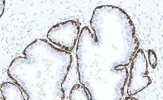
 Highly Definitive Antibodies - A range built around your IHC needs We analyzed over 60,000 points of data from laboratory staff and pathologists world-wide, to provide a range of high quality antibodies for your IHC needs.
 -
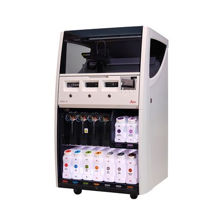
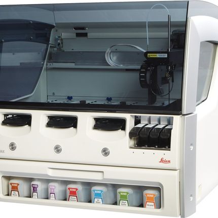 Bond MAX – Highest Quality, Automated, Economical, IHC - ISH Staining Available. Leica Bond MAX with 3 independent slide trays and easy reagent access.
 -
 Bond III Highest Productivity IHC- ISH staining available Bond III Showing intelligent reagent status with color coded illumination.
 -
 DS9800 Bond Polymer Refine Detection
 -
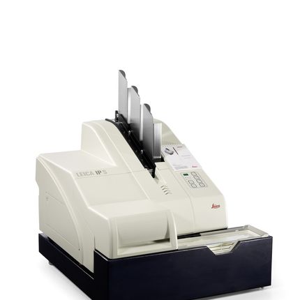 The Leica IP S imprints standard microscope slides, taking only four seconds per imprint when running in serial mode. A patented ink, specially designed for Leica Microsystems, makes imprints resistant to chemical exposure and physical wear. Whether alphanumeric characters, barcodes or logos, the print resolution is always excellent with good legibility. A specific advantage of barcode imprints is the tracking possibility of the probe within the complete histology workflow and additionally the reliable archiving with quick and accurate case identification. An optional external magazine is available to pre-load slides.
 -
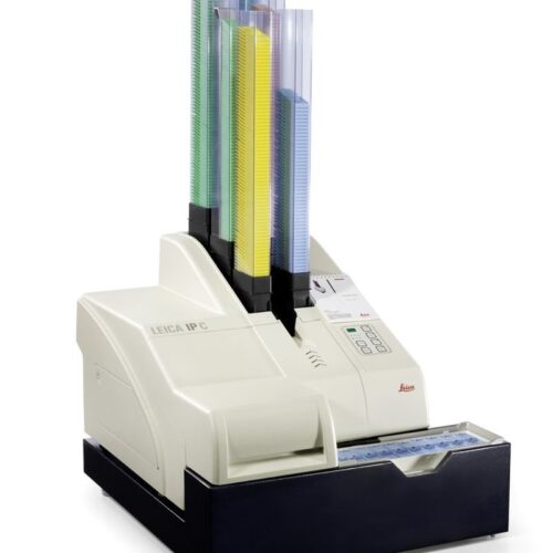 The Leica IP C has been designed for versatile printing of tissue cassettes, including cassettes with lids and two different imprint angles.
 -
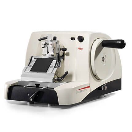
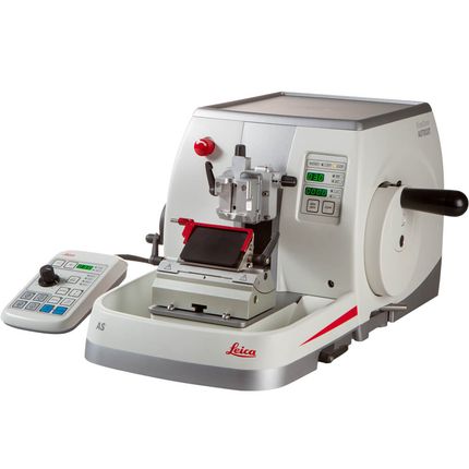 The new generation of microtomes from Leica Biosystems is built upon our 145+ year heritage of market-leading microtome design. It allows users to select among automated, semi auto-automated, or manual sectioning modes based their personal preferences.
 -
 The new generation of microtomes from Leica Biosystems is built upon our 145+ year heritage of market-leading microtome design. With new options such as the personalized coarse feed wheel and 2-in-1 blade holder, it ranks among the most ergonomically-friendly microtomes on the market. Quickly and efficiently select the turn direction most comfortable for you with the Personalized Coarse Feed Wheel.
 -
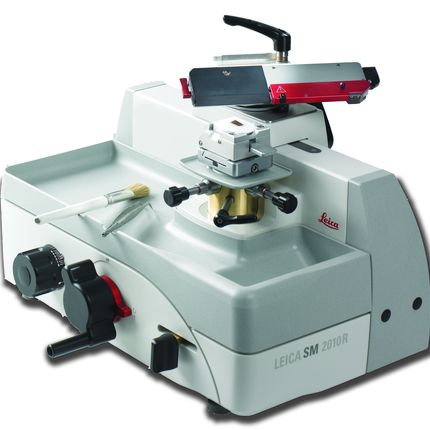 With smooth movement of the sledge, precision specimen orientation, and emphasis on safety and ergonomics, the SM2010 R produces high quality sections for routine histopathology (paraffin). The stable sliding microtome has a totally enclosed micrometer feeding system with an ergonomically positioned object head close to the user. The smooth-running sledge can be locked in 11 positions by using the easily accessible sledge brake.
 -
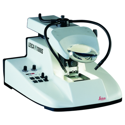 The Leica VT1000 S vibrating blade microtome is the instrument of choice for high-quality sectioning requirements in neurophysiology, neuropathology experimental pathology), Botany (roots and plants) and Industry (foams).
 -
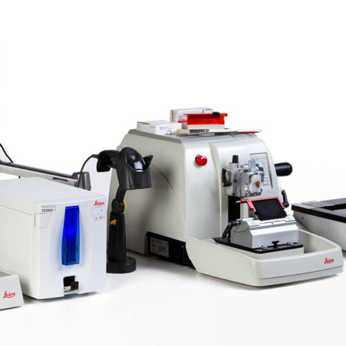 The Compact Slide Printing Solution that helps to reduce errors and maintain efficiency. We know how important patient safety is to your laboratory. With the PERMA S slide printer solution you can print individual slides at any microtome workstation. This provides the confidence that you have the right patient tissue associated with the right case number. On-demand direct printing helps reduce the risk of specimen misidentification and securely links patients to samples. The HistoCore PERMA S slide printer, combined with the validated PERMASLIDE slides, generates the high-quality printed barcodes required in your laboratory.
 -
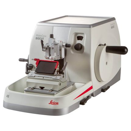
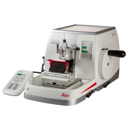 When you need the precision and speed of manual sectioning with the efficiency benefits of a semi-automated setup, the HistoCore MULTICUT saves the day. The new generation of microtomes from Leica Biosystems is built upon our 145+ year heritage of market-leading microtome design. Combining the speed of a manual microtome with the precision of motorized specimen feeding, MULTICUT helps you get consistent, reproducible sections. Produce high quality sections for IHC when using MULTICUT with the RM CoolClamp.
 -
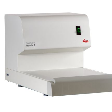 HistoCore Arcadia C is a cold plate holding more than 60/65 cassettes on its large working surface. Cooling efficiency is important, so the cold plate was designed with an environment adaptive control module to make sure the operating temperature is always stabilized at -6 °C. There is no need to turn down the temperature in summer or worry about too fast cooling in winter. The Arcadia C cold plate can be used as a stand-alone unit for re-cooling blocks prior to sectioning or as part of an embedding center to cool molds after paraffin dispensing.
 -
 The RM2125 RTS manual microtome has the essential features you need for safe, economic sectioning and optimized workflows.
 -
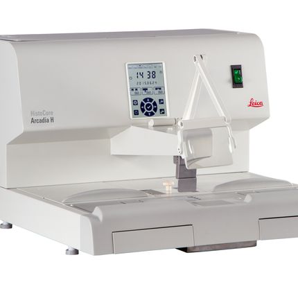 We know how important embedding is for you, so we designed the HistoCore Arcadia H Paraffin Dispenser to meet your needs. The HistoCore Arcadia H allows for simple operation and precise control, resulting in improved quality, a smooth workflow, reliability and speed of your embedding work. Features include wristpads for increased comfort and stability; an improved magnifier for small biopsy embedding; and an LCD touchscreen for increased control and monitoring of the instrument.
 -
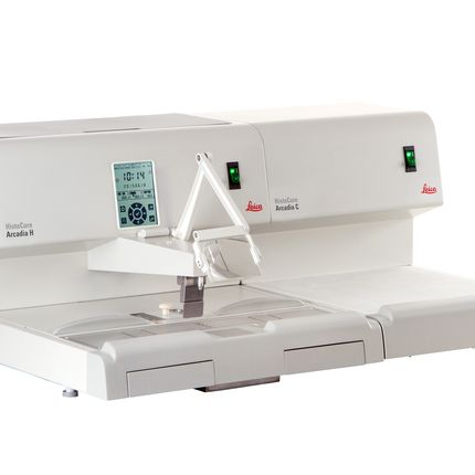 The HistoCore Arcadia modular tissue embedding system incorporates two separate components, the Arcadia H heated embedding module and the Arcadia C cold plate. The independent modules offer the flexibility to arrange the embedding workflow in the direction that best suits your laboratory.
 -
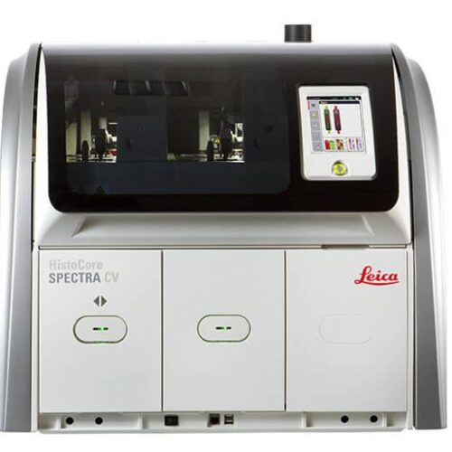
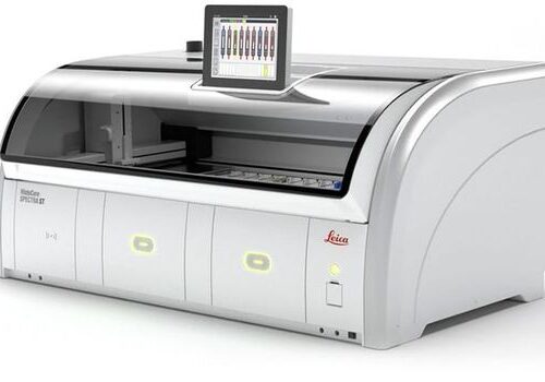 The HistoCore SPECTRA ST is a high throughput routine H&E stainer with the uncompromised flexibility to process H&E and special stains simultaneously by maintaining high throughput. The optimized and validated protocols with specifically designed consumables provide unmatched and consistent staining results for the wide range of intensities pathologists expect.
 -
 The Leica CV5030 fully automated glass coverslipper produces slides with superior optical quality for reliable long-term storage. Its capability of handling a large variety of slide racks from different suppliers makes the Leica CV5030 very flexible. Most common mounting media, including xylene-free varieties, can be used. The operator can choose wet or dry coverslipping. The CV5030’s performance achieves three goals: Highly consistent and reliable coverslipping quality, full adaptation to individual laboratory set-ups, integration into staining/coverslipping workstations to form a fully automated, user-friendly operation system.
 -
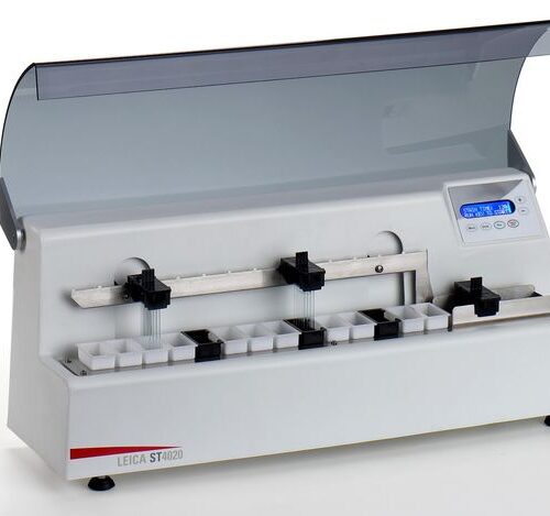 Leica ST4020 Small Linear Stainer delivers consistent quality in a compact design.
 -
 Premium specimen staining and coverslipping with an unmatched combination of ease of use, flexibility, productivity, and environmental friendliness Enjoy the benefit of LEAN workflows, achieved with Leica Microsystems' patented CodeRack™ technology.
 -
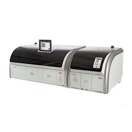 Your time matters. Speed up slide processing while maintaining quality and consistency with the HistoCore SPECTRA Workstation, the only workstation that provides consistent staining from slides 1 to 1,600. It also offers the fastest glass coverslipping drying time on the market at only 5 minutes, processing slides that are instantly dry to the touch.
 -
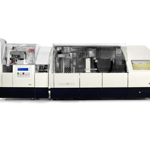
 The first and only solution in the market with dual glass coverslip lines enabling the highest throughput: up to 570 dried slides per hour. With an integrated hot air-drying oven, it achieves a drying time of only 5 minutes, providing the fastest turnaround time for your lab.
 -
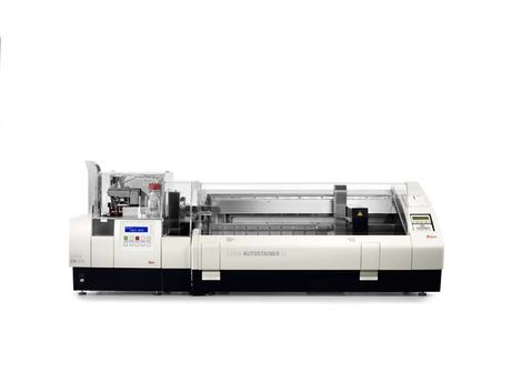
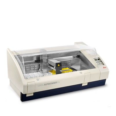 The proven Autostainer XL continues to provide reproducible, consistent, high-quality staining and increased workload throughput.
 -
 Increase your laboratory productivity with Leica Biosystems’ fully automated instruments providing high-quality staining, and superior glass coverslipping for routine H&E and Cytology.
 -
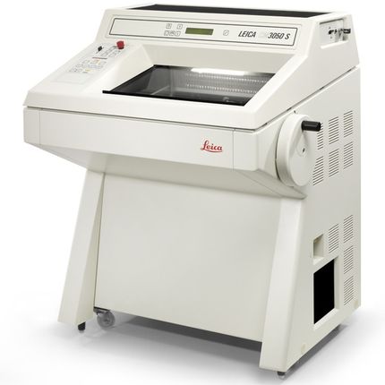 Primarily designed for the demanding needs of cryosectioning in biomedical, neuro-anatomical and pharmaceutical research. The Leica CM3050 S cryostat features superior user comfort with excellent safety standards for practically all types of cryosectioning applications. It is the instrument of choice for all research applications and for advanced clinical cryosectioning needs. Particularly when working with delicate specimens – for example brain samples in neuroscience – the precise specimen orientation and the specimen feed system guarantees reproducible, thin, serial sections of maximum quality.
 -
 The Leica CM1860 UV, developed with an emphasis on diagnostic confidence, safety and ergonomics, is the cryostat for histopathology laboratory applications. The high-throughput cryostat creates diagnostic confidence by reliably producing quality sections that help provide an accurate diagnosis.