-
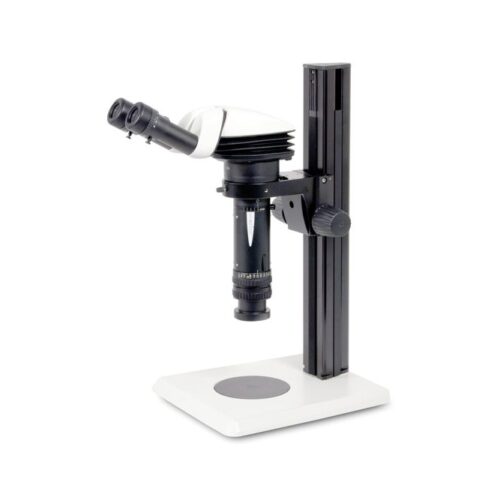
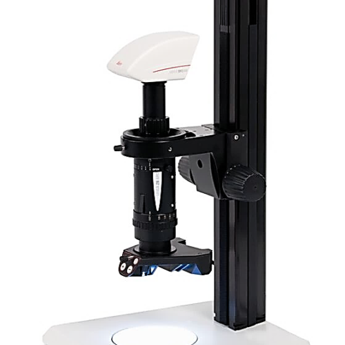 Manual 6:1 Macroscope for detailed Documentation, Measurement and Evaluation. The Leica Z6 APO is a fully apochromatic zoom system with excellent light transmission for high contrast, high-resolution, detailed analysis. The single beam path provides 2D images and ensures parallax-free imaging. the Leica Z6 APO with 6.3:1 zoom offers the highest numerical aperture: 0.117 nA (351 Lp/mm resolution) with the 1× plan-apochromatic objective
 -
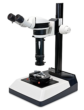 Leica Z16 APO features a 16:1 zoom, with a range from 0.57× to 9.2×. The high magnification Leica Z16 APO is suitable for deployment in microelectronics as well as laboratory workstations in medicine, biology, education, research, development, and forensics. With the standard configuration (apochromatic 1× objective and 10× eyepieces) objects can be observed at a magnification level of up to 115x and, depending on the optics combination, to a maximum of 2300x
 -
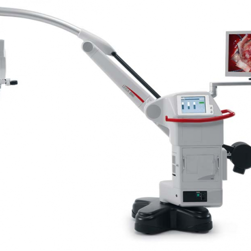
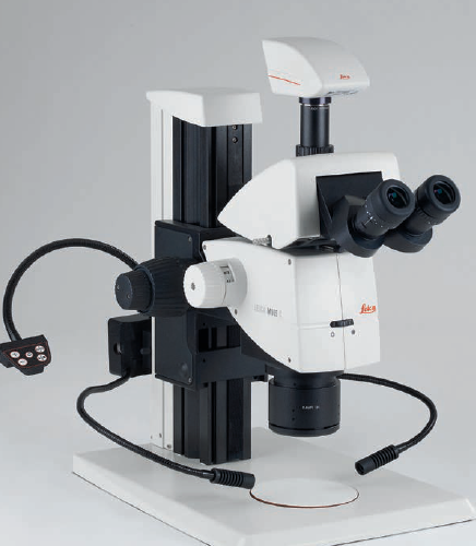
Neurosurgery Microscope
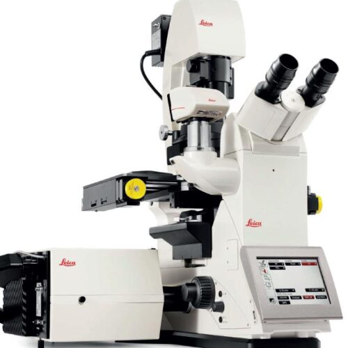
The Leica M530 OH6 takes image quality to a new level: FusionOptics, the groundbreaking technology by Leica Microsystems, unites an enhanced depth of field with high resolution to create an optimal view. FusionOptics also helps you to streamline your work: A larger area in full focus means that there is less need to refocus the microscope. FusionOptic let you stay focused – in every sense of the word. -
 The proprietary nature of the low-profile disposable blades DB80LX aids in cutting high quality, thin sections with even very difficult to section tissue including dense specimens. With select materials, sophisticated coating and an advanced profile, this blade quickly and efficiently allows you to section routine specimens, biopsies and dense tissues.
 -
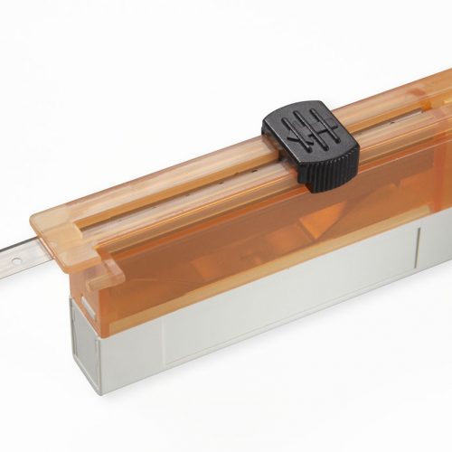
 Cut the very thin sections needed for diagnostic clarity. The low-profile disposable blades DB80LS provide a new level of quality with a low-profile shape, a premium finish and edge-to-edge, blade-to-blade consistency.
 -
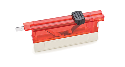
 Low-profile 819 disposable blades are 80 mm long x 8 mm high x 0.25 mm thick. Made of stainless steel, the blades are ultra-fine yet durable and specifically designed for use with Leica Biosystems microtomes or cryostats classified and labelled as in vitro diagnostic medical devices for histological medical diagnosis, e.g. cancer diagnosis.
 -
-
-
 The Leica Z6 APO is a fully apochromatic zoom system with excellent light transmission for high contrast, high-resolution, detailed analysis. The single beam path provides 2D images and ensures parallax-free imaging.
 -
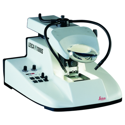 The Leica VT1000 S vibrating blade microtome is the instrument of choice for high-quality sectioning requirements in neurophysiology, neuropathology experimental pathology), Botany (roots and plants) and Industry (foams).
 -
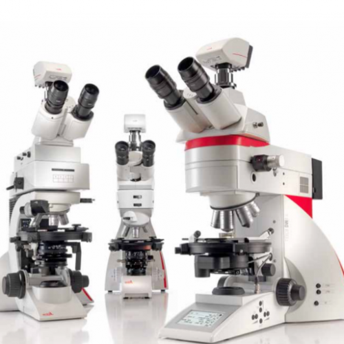 Depending on your application, you can choose from three different systems for polarization microscope. The Leica polarization microscope series is designed for all polarizing examinations: petrography, mineralogy, structure characterization, asbestos analysis, coal analysis (vitrinite reflection), and examination of liquid crystals.
 -
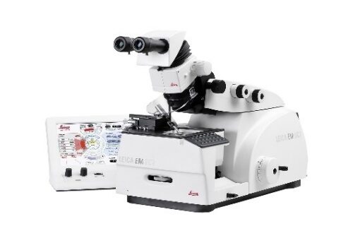 The Leica EM UC7 prepares excellent quality semi- and ultra-thin sections, as well as the perfectly smooth surfaces required for LM, TEM, SEM, and AFM examination. The precision mechanics, ergonomic design, and intuitive layout of the touchscreen control unit make the Leica EM UC7 ideal for the highest quality specimen preparation
 -
 The Leica TCS SPE is an affordable entry to the Leica family of confocals. It combines a straightforward design with high-end, true point-scanning confocal technology.
 -
 The Leica TP1020 tissue processor is available in four configurations: the basic instrument, the basic instrument with vacuum, the basic instrument with a fume control system and the basic instrument with both vacuum and fume control. Gentle specimen processing and a high level of specimen safety at all stages of the processing run are supported by the robust design based on precision mechanics in conjunction with a modern user interface.
 -
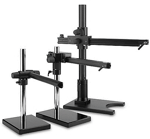 Leica Swingarm Stands (boom stands) are ideal to handle large samples. The high modularity allows to customize these stands depending on sample size and weight of the configuration. Focus arms and drives provide multiple mounting options and more safety features.
 -
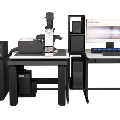 STELLARIS has been designed from the ground up to give you the power to see more. The synergy between the new Power HyD detectors family, the completely optimized beam path, and the new generation White Light Lasers (WLL) empowers you to obtain more accurate and reliable data to test your hypothesis with precision.
 -
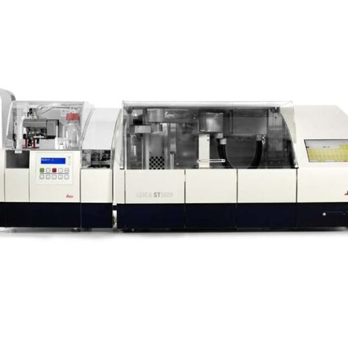 Premium specimen staining and coverslipping with an unmatched combination of ease of use, flexibility, productivity, and environmental friendliness Enjoy the benefit of LEAN workflows, achieved with Leica Microsystems' patented CodeRack™ technology.
 -
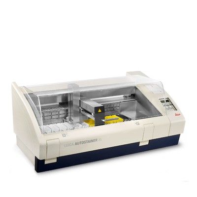
 Increase your laboratory productivity with Leica Biosystems’ fully automated instruments providing high-quality staining, and superior glass coverslipping for routine H&E and Cytology.
 -
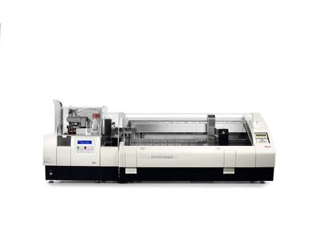
 The proven Autostainer XL continues to provide reproducible, consistent, high-quality staining and increased workload throughput.
 -
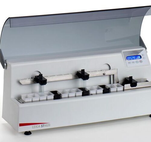 Leica ST4020 Small Linear Stainer delivers consistent quality in a compact design.
 -
 The Leica LED3000 SLI and Leica LED5000 SLI each feature two bright LED spotlights, which can be individually adjusted to the sample with two goosenecks. The control element is mounted on a separate gooseneck and can be placed in any position.
 -
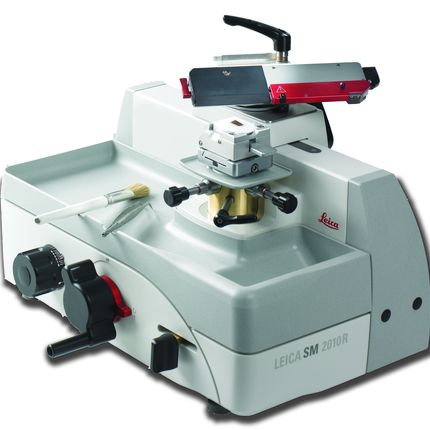 With smooth movement of the sledge, precision specimen orientation, and emphasis on safety and ergonomics, the SM2010 R produces high quality sections for routine histopathology (paraffin). The stable sliding microtome has a totally enclosed micrometer feeding system with an ergonomically positioned object head close to the user. The smooth-running sledge can be locked in 11 positions by using the easily accessible sledge brake.
 -
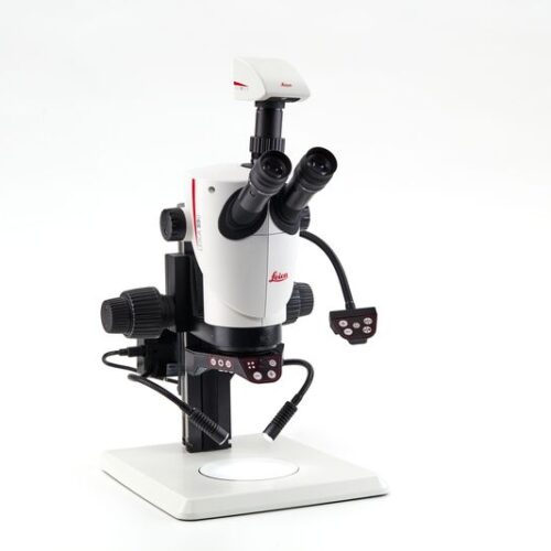
 Enjoy working with HD-microscope images live streamed to your PC, HD-monitor, or mobile device. The S9 i stereo microscope has an integrated 10 MP CMOS-camera and can be connected with your facility’s network by Ethernet. This gives you the opportunity to quickly react to queries, get a second opinion, and discuss problems e.g. via tablet with others.
 -
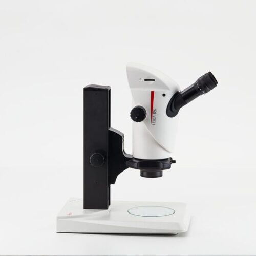
 The S9 D gives you the flexibility to add a camera to the microscope and turn it into a digital microscope solution. The built in 50:50 split of the beam path allows observation through the eyepieces and digital imaging at the same time.