-
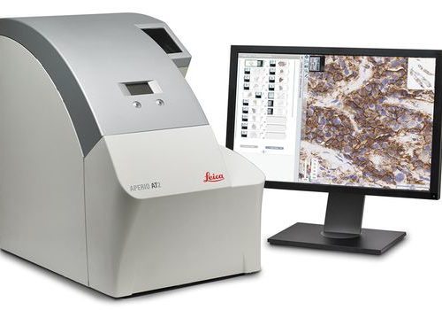 With its compact size, high capacity autoloader and optimal image quality, the Aperio AT2 slide scanner has set new standards in digital pathology. Aperio AT2 consistently delivers precise, whole slide images with an unparalleled, low rescan rate. Slides are available for remote viewing in less than a minute. With an easy-to-use Pathologist's cockpit, coupled with easy integration into laboratory information systems (LIS), the Aperio AT2 provides an ideal platform for research institutions.
 -
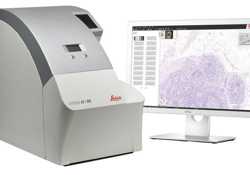 ntroducing the Aperio AT2 DX System, a 400 slide scanner with Aperio ImageScope DX clinical viewing software, and a high-performance viewing workstation including color-calibrated medical grade monitors. The system can be used standalone, or in conjunction with Aperio Path DX case management software, for a streamlined, clinical workflow. Developed with proven technology from Leica Biosystems, the Aperio AT2 DX System enables high volume clinical labs to produce consistent, diagnostic quality images, enabling pathologists to diagnose with confidence.
 -
 Create high-quality digital slides with the ultra-compact Aperio CS2 image capture device from your desktop. With a five-slide capacity and 20X and 40x magnification capabilities, the Aperio CS2 is a highly reliable workhorse for medium-volume sites.
 -
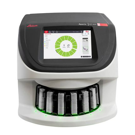 The Aperio GT 450 enables histotechnicians to complete scanning tasks quickly and with confidence leveraging a 32-second scan speed*. Output 81 slides/hr at 40x* delivering high quality images with Leica optics and with an IT architecture that is secure and scalable. From the pathology lab to the IT room, the Aperio GT 450 is designed to scale up digital pathology operations.
 -
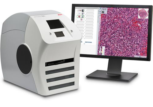
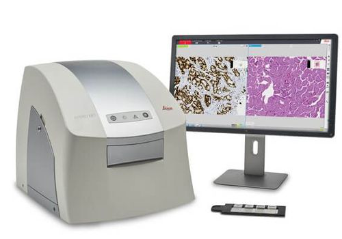 The Aperio LV1 is a cost-effective solution for research users looking to adopt Digital Pathology. The small footprint and compact design makes the Aperio LV1 an ideal desktop system. With minimal IT requirements and rapid implementation, your Aperio LV1 can be installed in minutes.
 -
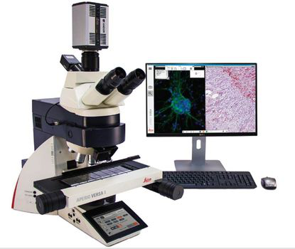 The Aperio VERSA is a comprehensive digital pathology scanner, designed and developed to support the diverse imaging needs of cutting edge research facilities. The combination scanner is optimized for precision scanning of brightfield and fluorescent samples, with the accuracy and resolution required for FISH.
 -
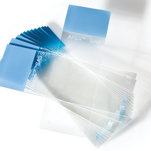 The Apex Adhesive Slides portfolio consists of microscope slides made of white glass and coated with a proprietary adhesive coating for a positive charge, to promote tissue adhesion and to allow the slide to be more hydrophilic.
 -
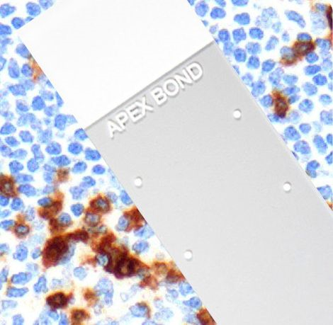 The Apex BOND is an extension of the Apex Adhesive Slides portfolio that is specifically designed and validated for IHC staining on the BOND platform. The eight dots represent the usable areas of the Apex BOND for the different dispense volumes in the BOND processing modules.
 -
 The Apex clipped corners are clipped at 45° angles to help reduce glass breakage and fragmentation when loaded into slide printers.
 -
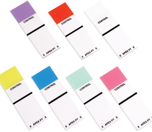 Apex Patient Control Box Slides have clearly designated areas for positive control and patient tissues.
 -
 Beveled Edge Slides feature beveled edges and corners that make for easier handling. The 25 mm x 75mm slides are washed and carefully inspected to ensure quality.
 -
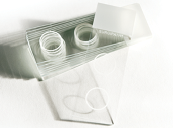 These slides are 3”x 1” White Snowcoat slides with two circles, each having a diameter of 1/2”. These circles match up with the Cytology Funnel, which is to be used for preparation of non-gyn cytology specimens.
 -
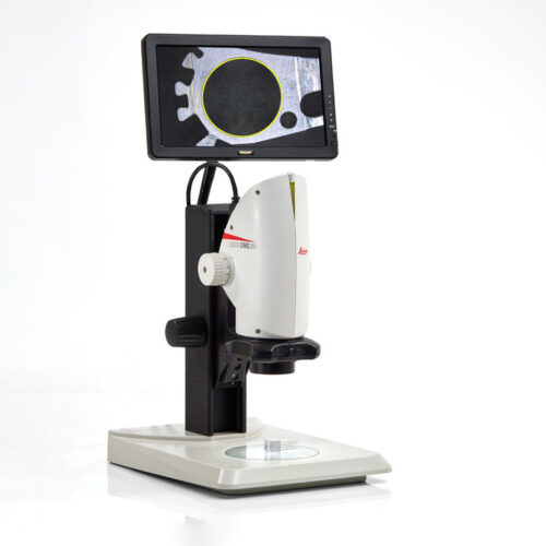 Digital microscope system for digital inspection, observation and measurement. From tiniest detail to an overview, the optics ensures magnification of up to 300x. The built-in HDMI microscope camera provides full high definition live images of up to 30fps and a resolution of 5Mpixels.
 -
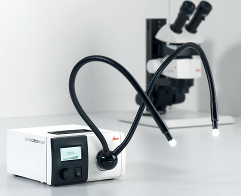 The Leica CLS LED light source has been developed for routine and high-end microscopy applications. It is compatible with all fiber-optic light guides of the LED series.
 -
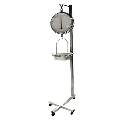 These absolute precision scales have a large 13 inches (330 mm) double dial with chrome-plated housing. The scales have large numerals and graduations for easy reading. The removable pan is constructed of stainless Steel and features drain holes
 -
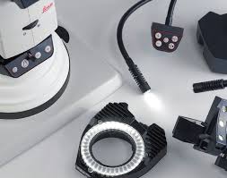
 The Leica LED5000 / Leica LED3000 family is also fully integrated into the complete system of stereomicro¬scopes and accessories from Leica Microsystems Focusing columns with integrated electronics connect all digital signals and send them to Leica Application Suite software.
 -
 The Leica LED5000 / Leica LED3000 family is also fully integrated into the complete system of stereomicroscopes and accessories from Leica Microsystems Focusing columns with integrated electronics connect all digital signals and send them to Leica Application Suite
 -
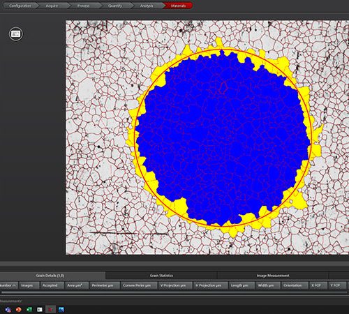 For industrial quality control and materials research, your goal is to deliver accurate and reliable results. You need software that enables you to work with speed, efficiency, and precision and facilitates your daily tasks.
 -
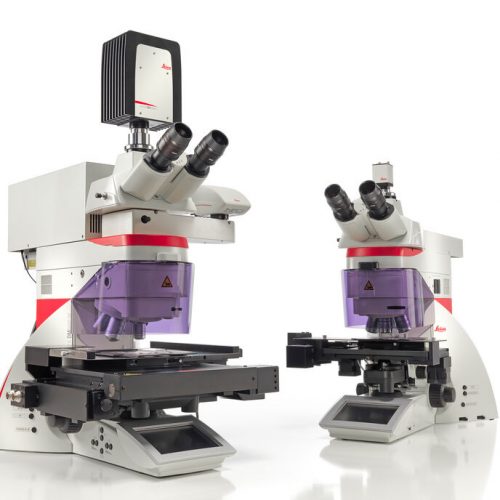 Laser Microdissection (LMD, also known as Laser Capture Microdissection or LCM) enables users to isolate specific single cells or entire areas of tissue. Powered by a unique laser design and dynamic software, Leica LMD systems allow users to easily isolate Regions of Interest (ROI) from entire areas of tissue down to single cells or even subcellular structures such as chromosomes.
 -
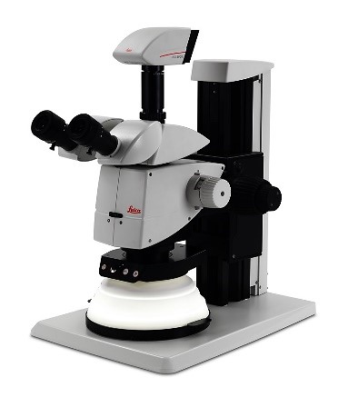
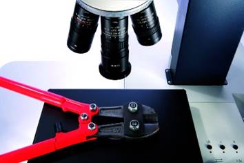 The intuitive A60 F and A60 S stereo microscopes fulfill what you need – high sample throughput, optimum visibility of product details and components, and easy processing of subassemblies.
 -
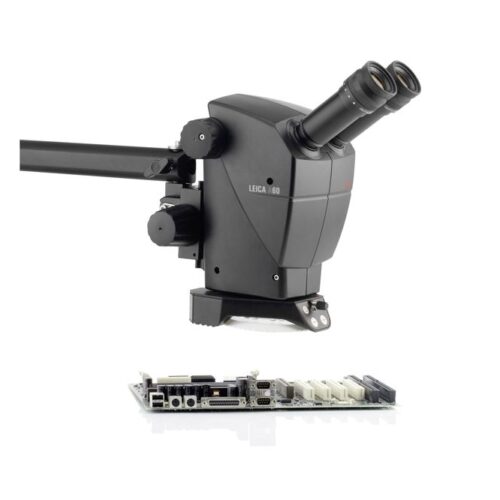 The intuitive A60 F and A60 S stereo microscopes fulfill what you need – high sample throughput, optimum visibility of product details and components, and easy processing of subassemblies. The Leica A60 stereomicroscope does not require the user to be a microscope expert. The components of the preconfigured system are perfectly matched to each other to provide easy operation with the best optical results. Choose between two different stands: the Leica A60 F with flex arm for maximum freedom of movement or the Leica A60 S with swing arm for high stability.
 -
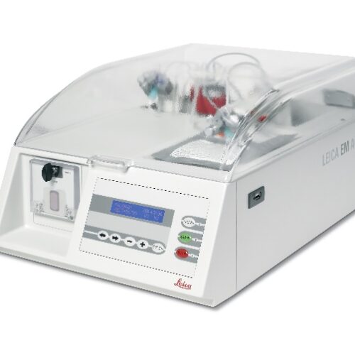 The Leica EM AC20 automatic contrasting system for ultrathin sections ensures minimum user contact with reagents and reduced reagent consumption.
 -
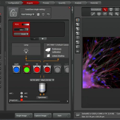 The Leica Application Suite (LAS) integrates Leica automated microscopes and digital cameras and provides one common, easy-to-use, consistent user interface.
 -
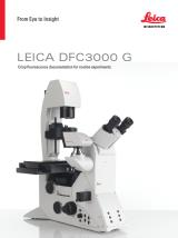 The Leica DFC3000 G is a grayscale USB 3.0 microscope camera for routine fluorescence applications. You will receive crisp images due to its unique passive cooling architecture and its highly sensitive CCD sensor. Correlated pixel double sampling together with a reduction of the ambient temperature of the sensor offers an exceptionally clear and noise -free signal. Its CCD sensor is particularly suitable for low light situations such as fluorescence and will capture even smallest amounts of light.
 -
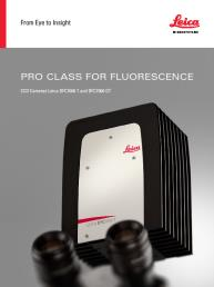 These cooled 2.8 megapixel cameras provide a new paradigm for camera image quality through truly innovative design. The monochrome DFC7000 GT for demanding fluorescence applications, or the color DFC7000 T developed for both brightfield and fluorescence imaging.
 -
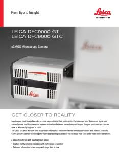 The Leica DFC9000 sCMOS camera will turn your imagination into reality. This monochrome microscope camera with newest scientific CMOS (sCMOS) sensor technology for fluorescence imaging enables you to image your cells under near-native conditions.
 -
 Fluorescence is one of the most commonly used physical phenomena in biological and analytical microscopy, mainly because of its high sensitivity and high specificity. Fluorescence is a form of luminescence.
 -
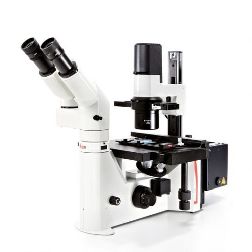
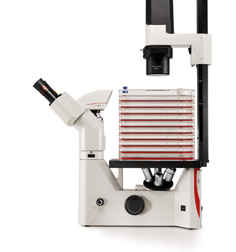 The Leica DM IL LED features a comprehensive set of contrast methods to monitor your specimen the way you need. High-quality Phase contrast, excellent modulation contrast and brilliant fluorescence are just one fingertip away. Robust stability, plenty of space to work with tools, long working distances to accommodate large culture flasks and a stable illumination without heat make work at the microscope easy and convenient.
 -
 Fluorescence is one of the most commonly used physical phenomena in biological and analytical microscopy, mainly because of its high sensitivity and high specificity. Fluorescence is a form of luminescence.
 -
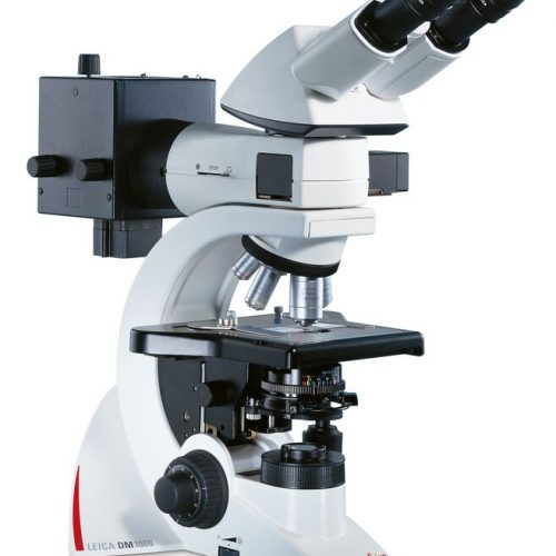
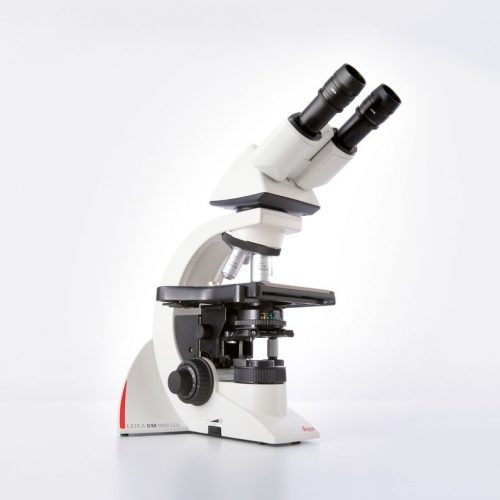 The Leica DM1000 LED features long-life LED illumination that provides near daylight, bright illumination with constant colour temperature and emits less heat.
 -
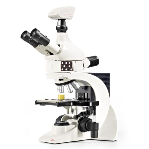 The Leica DM1750 M is a material microscope designed for rapid, accurate analysis results even for a use in rough ambient conditions. Working with the Leica DM1750 M you will see; how simple and reliable microscopy can be. Its robust design contains an excellent optical system and allows the inspection even of larger samples, in brightfield, oblique- or with polarized light. The entire reflected light illumination is carried out with Power-LEDs which allow an inspection with different illumination angles, especially suitable for the detection of micro scratches or for gaining height information.
 -
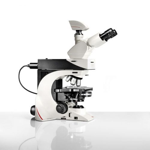
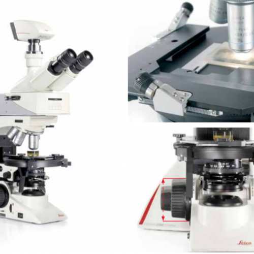 The Leica DM2700 M flexible upright microscope system uses LED illumination for all contrast methods: brightfield (BF), darkfield (DF), differential interference contrast (DIC), qualitative polarization (POL), and fluorescence (FLUO) applications. It also offers built-in oblique illumination, which improves the visualization of surface topography and defects. Optionally, the Leica DM2700 M can also be equipped with a transmitted light axis. The Leica DM2700 M is equipped, e.g. with an N PLAN achromatic objective series with magnifications from 5x to 100x, a field of view of 22 mm, a flattened image field, and large working distances.
 -
 Fluorescence is one of the most commonly used physical phenomena in biological and analytical microscopy, mainly because of its high sensitivity and high specificity. Fluorescence is a form of luminescence.
 -
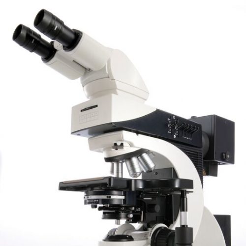
 With their sophisticated modular design and high-performance fluorescence, the Leica DM2000 microscopes are ideal for complex tasks in pathology, cytology, and many other applications. For special diagnostics requirements, the microscope is certified for in-vitro-diagnostics (IVD) like in-vitro-fertilization (IVF).
 -
Fluorescence is one of the most commonly used physical phenomena in biological and analytical microscopy, mainly because of its high sensitivity and high specificity. Fluorescence is a form of luminescence.
-
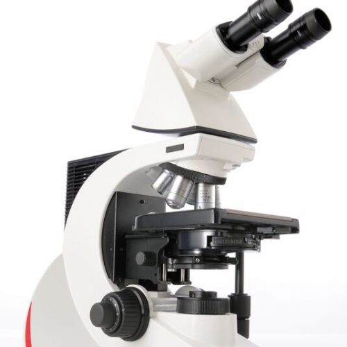
 Leica DM2500 & DM2500 LED optical microscopes are tools for demanding tasks in life science routine and research applications. With their transmitted light illumination, optical performance, and state-of-the-art accessories, they are especially well-suited for challenging life science research tasks that require differential interference contrast or high-performance fluorescence.