-
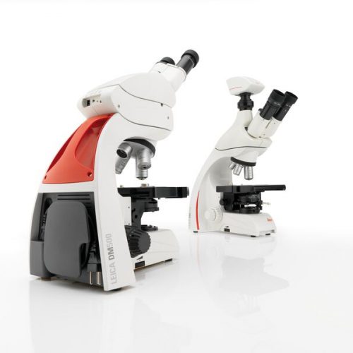 The Leica DM500 microscope with “plug and play” capability is the ideal tool to make teaching entry-level college and university Life Science courses easy and fun for the instructor and the student.
 -
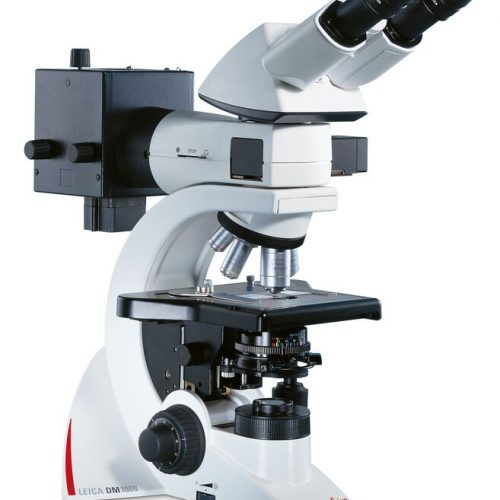
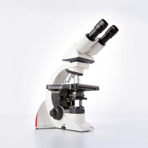 The Leica DM1000 LED features long-life LED illumination that provides near daylight, bright illumination with constant colour temperature and emits less heat.
 -
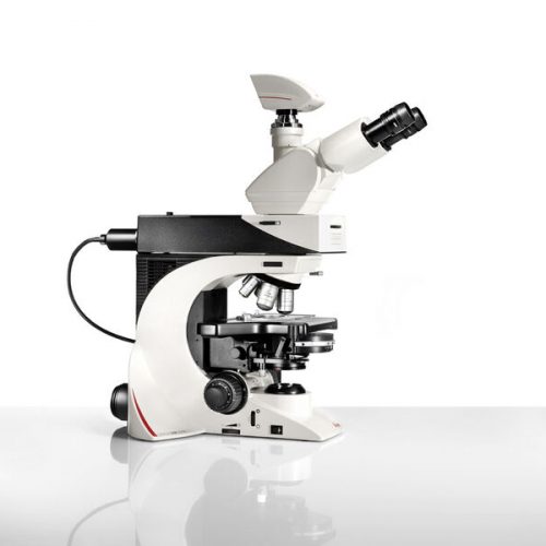
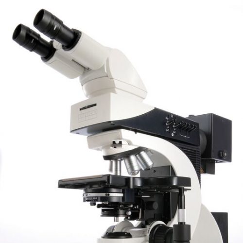
 With their sophisticated modular design and high-performance fluorescence, the Leica DM2000 microscopes are ideal for complex tasks in pathology, cytology, and many other applications. For special diagnostics requirements, the microscope is certified for in-vitro-diagnostics (IVD) like in-vitro-fertilization (IVF).
 -
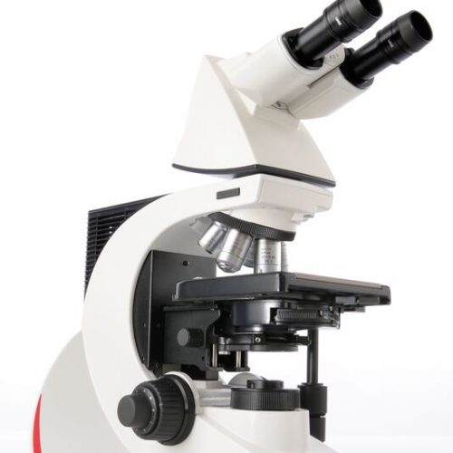
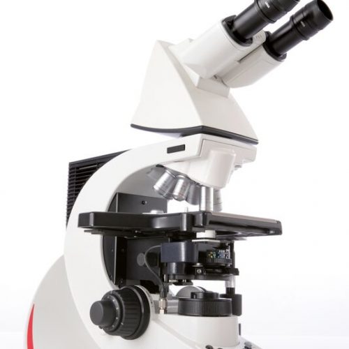
 Leica DM2500 & DM2500 LED optical microscopes are tools for demanding tasks in life science routine and research applications. With their transmitted light illumination, optical performance, and state-of-the-art accessories, they are especially well-suited for challenging life science research tasks that require differential interference contrast or high-performance fluorescence.
 -
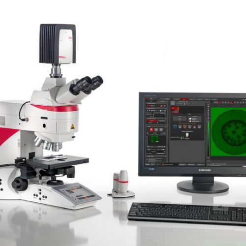
 The Leica DM3000 microscopes for pathology, cytology, haematology and many other applications feature a motorized nosepiece, condenser head, automated light intensity adjustment, and optional foot pedal. These intuitive microscopes improve workflows significantly. For special diagnostics requirements, the microscope is certified for in-vitro-diagnostics (IVD) like in-vitro-fertilization (IVF).
 -
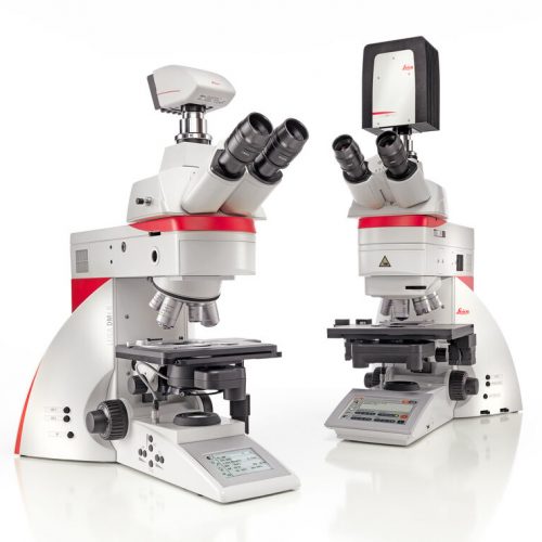 Increase your work efficiency with the Leica DM4 B or Leica DM6 B upright digital research microscopes! Both are ideally suited to making your work life easier in biomedical research and clinical labs alike. You can configure the microscope that fits your need and budget with the Leica DM4 B and Leica DM6 B. Optimize your system to your application with different contrast methods and automated functions.
 -
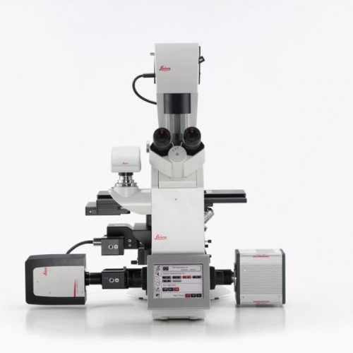
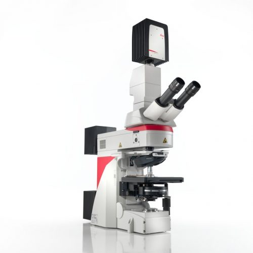 Do you work in a field such as electrophysiology, evolutionary biology, or neuroscience where outstanding stability is required for successful experiments? The Leica DM6 FS fixed stage fluorescence microscope is a suitable instrument for your present and future challenges.
 -
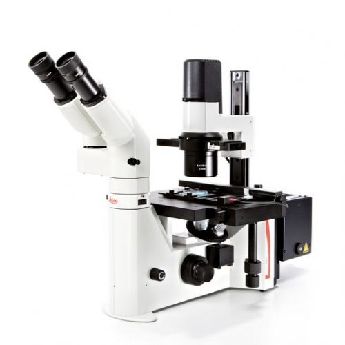
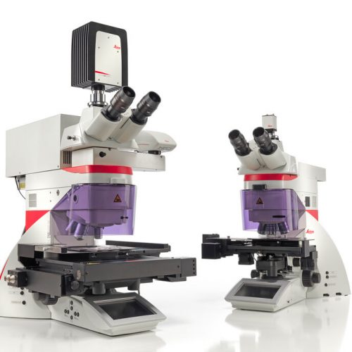 Laser Microdissection (LMD, also known as Laser Capture Microdissection or LCM) enables users to isolate specific single cells or entire areas of tissue. Powered by a unique laser design and dynamic software, Leica LMD systems allow users to easily isolate Regions of Interest (ROI) from entire areas of tissue down to single cells or even subcellular structures such as chromosomes.
 -
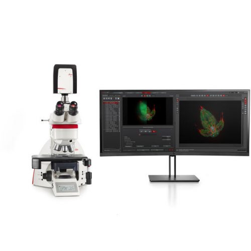 The THUNDER Imager Tissue allows real-time fluorescence imaging of 3D tissue sections typically used in neuroscience and histology research. Acquire rich, detailed images of thick tissues free of haze from out-of-focus blur.
 -
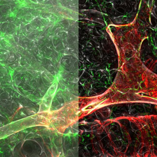
 Fluorescence is one of the most commonly used physical phenomena in biological and analytical microscopy, mainly because of its high sensitivity and high specificity. Fluorescence is a form of luminescence.
 -
 Fluorescence is one of the most commonly used physical phenomena in biological and analytical microscopy, mainly because of its high sensitivity and high specificity. Fluorescence is a form of luminescence.
 -
Fluorescence is one of the most commonly used physical phenomena in biological and analytical microscopy, mainly because of its high sensitivity and high specificity. Fluorescence is a form of luminescence.
-
 Fluorescence is one of the most commonly used physical phenomena in biological and analytical microscopy, mainly because of its high sensitivity and high specificity. Fluorescence is a form of luminescence.
 -
 Fluorescence is one of the most commonly used physical phenomena in biological and analytical microscopy, mainly because of its high sensitivity and high specificity. Fluorescence is a form of luminescence.
 -
 Fluorescence is one of the most commonly used physical phenomena in biological and analytical microscopy, mainly because of its high sensitivity and high specificity. Fluorescence is a form of luminescence.
 -
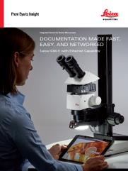
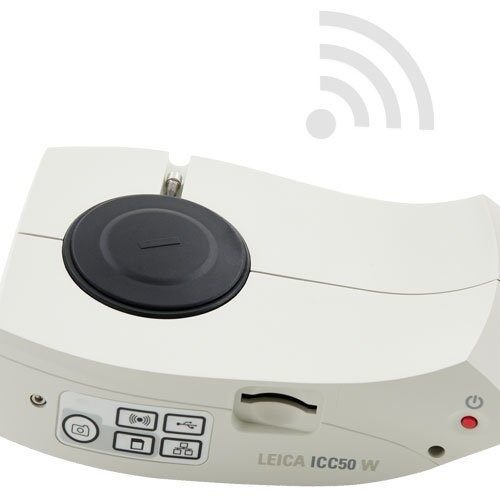 The ICC50 E exclusively uses the facilities network (WLAN or LAN) to allow students to connect to the camera. This is an ideal solution if you don`t want to add additional Wi-Fi access points to your existing wireless network concept. These 5-megapixel cameras can live-stream HD images to students’ smartphones or tablets. Students can connect to the ICC50 W either through their own internal Wi-Fi signal or through the facilities network.
 -
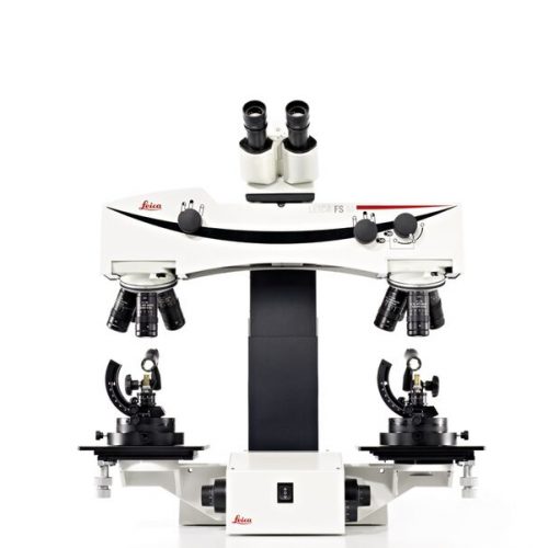 The Leica FS M manually-controlled forensic macroscope provides the flexibility, convenience, and comfort that make it the universal instrument of firearms and tool mark examiner. The versatile system is ideal for the simultaneous observation of evidence for training and consultation. The high-quality comparison bridge supports two precisely matched sets of apochromatically corrected objectives on quintuple ball-bearing nosepieces.
 -
 The time has come for imaging systems that allow you to tackle easily biologically relevant 3D models: THUNDER Imagers. To answer important scientific questions, they enable you to obtain a clear view of details, even deep within an intact sample, in real-time without out-of-focus blur.
 -
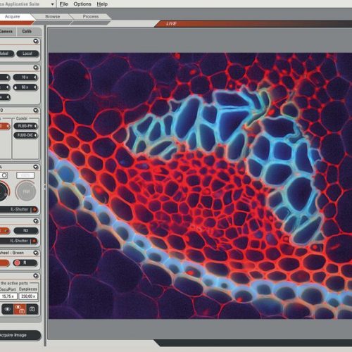
 The Leica Application Suite (LAS) integrates Leica automated microscopes and digital cameras and provides one common, easy-to-use, consistent user interface.
 -
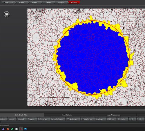 For industrial quality control and materials research, your goal is to deliver accurate and reliable results. You need software that enables you to work with speed, efficiency, and precision and facilitates your daily tasks.
 -
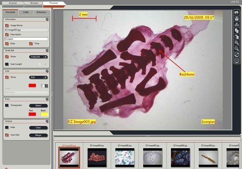 The Leica LAS EZ software, compatible with the Leica EZ4 W/E stereomicroscope, as well as the Leica EC4, Leica ICC50 W/E and Leica IC90 E digital cameras, provides an ideal common, easy-to-use, consistent platform for basic education, industry and life science applications.
 -
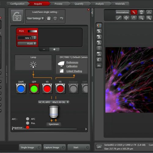
 Leica Application Suite X (LAS X) is the one software platform for all Leica microscopes: It integrates confocal, widefield, stereo, super-resolution, and light-sheet instruments from Leica Microsystems.
 -
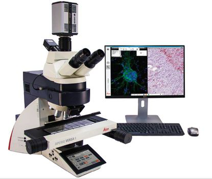 The Aperio VERSA is a comprehensive digital pathology scanner, designed and developed to support the diverse imaging needs of cutting edge research facilities. The combination scanner is optimized for precision scanning of brightfield and fluorescent samples, with the accuracy and resolution required for FISH.
 -
 Create high-quality digital slides with the ultra-compact Aperio CS2 image capture device from your desktop. With a five-slide capacity and 20X and 40x magnification capabilities, the Aperio CS2 is a highly reliable workhorse for medium-volume sites.
 -
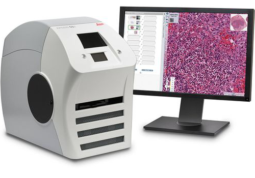
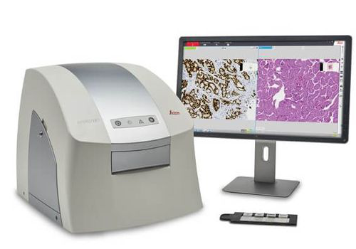 The Aperio LV1 is a cost-effective solution for research users looking to adopt Digital Pathology. The small footprint and compact design makes the Aperio LV1 an ideal desktop system. With minimal IT requirements and rapid implementation, your Aperio LV1 can be installed in minutes.
 -
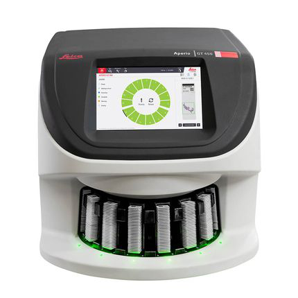 The Aperio GT 450 enables histotechnicians to complete scanning tasks quickly and with confidence leveraging a 32-second scan speed*. Output 81 slides/hr at 40x* delivering high quality images with Leica optics and with an IT architecture that is secure and scalable. From the pathology lab to the IT room, the Aperio GT 450 is designed to scale up digital pathology operations.
 -
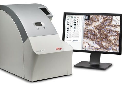 With its compact size, high capacity autoloader and optimal image quality, the Aperio AT2 slide scanner has set new standards in digital pathology. Aperio AT2 consistently delivers precise, whole slide images with an unparalleled, low rescan rate. Slides are available for remote viewing in less than a minute. With an easy-to-use Pathologist's cockpit, coupled with easy integration into laboratory information systems (LIS), the Aperio AT2 provides an ideal platform for research institutions.
 -
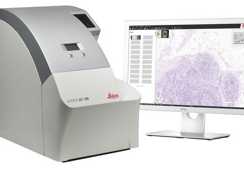 ntroducing the Aperio AT2 DX System, a 400 slide scanner with Aperio ImageScope DX clinical viewing software, and a high-performance viewing workstation including color-calibrated medical grade monitors. The system can be used standalone, or in conjunction with Aperio Path DX case management software, for a streamlined, clinical workflow. Developed with proven technology from Leica Biosystems, the Aperio AT2 DX System enables high volume clinical labs to produce consistent, diagnostic quality images, enabling pathologists to diagnose with confidence.
 -
 The Leica IC90 E CMOS microscope camera for stereo microscopes was developed for the Leica M series stereo microscopes. It provides pin-sharp images with spot-on color reproduction for professional visualization and documentation of industrial applications.
 -
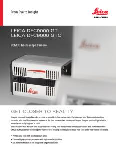 The Leica DFC9000 sCMOS camera will turn your imagination into reality. This monochrome microscope camera with newest scientific CMOS (sCMOS) sensor technology for fluorescence imaging enables you to image your cells under near-native conditions.
 -
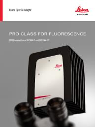 These cooled 2.8 megapixel cameras provide a new paradigm for camera image quality through truly innovative design. The monochrome DFC7000 GT for demanding fluorescence applications, or the color DFC7000 T developed for both brightfield and fluorescence imaging.
 -
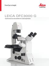 The Leica DFC3000 G is a grayscale USB 3.0 microscope camera for routine fluorescence applications. You will receive crisp images due to its unique passive cooling architecture and its highly sensitive CCD sensor. Correlated pixel double sampling together with a reduction of the ambient temperature of the sensor offers an exceptionally clear and noise -free signal. Its CCD sensor is particularly suitable for low light situations such as fluorescence and will capture even smallest amounts of light.
 -
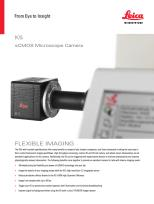 The K5 (grayscale) is ideal for routine fluorescence imaging applications such as immunostaining assays and 3D Cell Culture imaging. Image a wide variety of sample types with the flexibility and power of high 4.2megapixel resolution and fast 40 fps imaging brought to you by the K5.
 -
 The DMC6200 camera delivers amazing images with intense color detail and contrast - whether at the lowest or highest magnification. Its state-of-the-art CMOS sensor has a 5.86 μm pixel size, 2.3 megapixels sensor resolution, and an astounding dynamic range of 73 dB (4000:1). Achieve an image resolution of up to 20.7 megapixels with cutting-edge pixel shift technology.
 -
 The DMC5400 color CMOS camera offers high frame rate & high-resolution color images even at low magnification. It is optimized to produce fast, high quality images for documentation, evaluation and analysis, for a wide variety of samples and applications in manufacturing and life science research.
 -
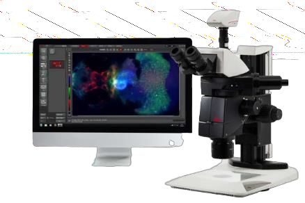
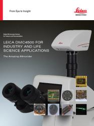 Leica DMC4500 color camera. It has been designed as a versatile, uncomplicated tool that simplifies the imaging process from capture through to processing. It is ideally suited to life science applications such as documenting slides and organisms, pathology or pharmaceutical testing and industrial applications such as quality control and failure analysis.